ความคิดเห็นทั้งหมด : 3

A 50-YOW had 6-month history of unilateral rhinorrhea.


   หญิงอายุ 50 ปี (หมดประจำเดือนแล้ว) ซึ่งมี asthma และ allergic rhinitis มาโรงพยาบาลเนื่องจากมีน้ำออกจากรูจมูกข้างขวามานาน 6 เดือน เมื่อตื่นนอนตอนเช้า ผู้ป่วยสังเกตว่ามีรอยเปื้อนที่หมอน และมีน้ำออกมาจากจมูกทุกครั้งเมื่อจามหรือไอ ไม่มีปวดหัว, น้ำหนักเปลี่ยนแปลง, น้ำนมไหลหรือหรือมองเห็นผิดปกติ เมื่อผู้ป่วยโน้มตัวไปข้างหน้า (เช่น เพื่อใส่รองเท้า) ก็จะมีน้ำใสๆ หยดออกมาจากรูจมูกข้างขวาหลายหยด

1. Unilateral rhinorrhea ในผู้ป่วยรายนี้น่าจะออกมาจากที่ไหน
และจะตรวจยืนยันได้อย่างไร
2. จะ manage อย่างไร


Posted by : cpantip , E-mail : (chpantip@medicine.psu.ac.th) ,
Date : 2010-03-25 , Time : 11:52:58 , From IP : 172.29.3.68


ความคิดเห็นที่ : 1


   ขอลองตอบครับ
-->ข้อ1.Unilateral rhinorrhea ในผู้ป่วยรายนี้น่าจะออกมาจากที่ไหน และจะตรวจยืนยันได้อย่างไร?
>สำหรับปัญหาเรื่อง unilateral rhinorrhea มี differential diagnosis ได้แก่ choanal atresia, nasal foreign body, nasal neoplasm, cerebrospinal rhinorrhea ทั้งนี้สองประการแรกนั้นมักตรวจพบในผู้ป่วยเด็กมากกว่า สำหรับสองประการหลังนี้อาจแยกเพียงตรวจโพรงจมูกดู อย่างไรก็ตามน้ำดังกล่าวไหลออกทางจมูกทุกครั้งเมื่อจามหรือไอ(เกี่ยวข้องกับการเพิ่ม ICP) หรือเวลาโน้มตัวไปข้างหน้า ทำให้นึกถึง CSF rhinorrhea ซึ่งเข้ากับประวัติดังกล่าวได้มากที่สุด หากนำรอยเปื้อนที่หมอนมาดู อาจจะพบ halo sign (รอยเลือดตรงกลางวงล้อมรอบด้วยรอยน้ำใส)
>สำหรับการตรวจยืนยันนั้น เนื่องจากสารคัดหลั่งอาจปนกับเลือดดังนั้นการส่ง Beta-2 transferrin ซึ่งเป็น carbohydrate-free isoform ของ transferring ซึ่งมีความจำเพาะหรือพบได้ใน CSF(sensitiviry ใกล้เคียง 100% และ specificity 95% โดย Beta-2 transferrin นี้พบได้เพียงใน perilymph, vitreous humor และ CSF เท่านั้น) โดยเลือดหรือ nasal secretion ไม่สามารถรบกวนผลตรวจได้
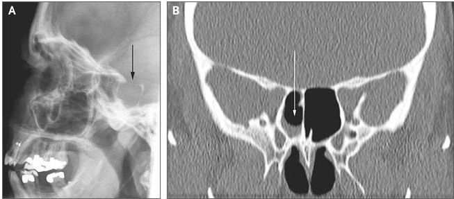
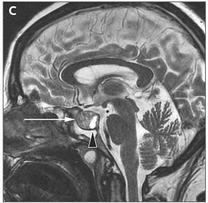
>สำหรับสาเหตุนั้น แม้ว่า traumatic leaks เป็นสาเหตุส่วนใหญ่กว่า 80% แต่ไม่มีประวัติอุบัติเหตุหรือผ่าตัด ดังนั้นอาจต้องคิดถึงกลุ่ม nontraumatic ได้แก่ brain tumors(intracranial และ extracranial tumor, cholesteatoma หรือ tuberculoma ซึ่งจะ erode กระดูกโดยตรง), skull base congenital defect และ meningoceles/meningoencephaloceles ในรายนี้คงต้องคิดถึง brain tumor เนื่องจากสาเหตุสองประการหลังมักตรวจพบในผู้ป่วยเด็ก
-->ข้อ2. จะ manage อย่างไร?
1.หลักการที่สำคัญของการรักษา CSF leakage คือ localization ของ dural defect โดยใช้ imaging ได้ CT/MRI
2.ให้ผู้ป่วยนอนบนเตียงยกหัวสูง, เลี่ยงการสูด/จาม/ไอ แรงๆ, ใช้ stool softeners
3.รอผล imaging เพื่อยืนยันการวินิจฉัยและดำเนินตามแนวทางการรักษาที่เหมาะสม
[ref: Abuabara A, Med Oral Patol Oral Cir Bucal. 2007 Sep 1;12(5):E397-400. Review.
Kerr JT, Otolaryngol Clin North Am. 2005 Aug;38(4):597-611. Review.]


Posted by : weeratian , Date : 2010-03-28 , Time : 20:07:02 , From IP : 112.142.28.229

ความคิดเห็นที่ : 2


   ในน้ำจากจมูกของผู้ป่วยมี β2-transferrin, ซึ่งเป็นการตรวจพบที่เข้าได้กับ cerebrospinal fluid (CSF).

Radiography of the sinuses revealed an enlarged sella turcica (Panel A, arrow), and a computed tomographic scan showed an air–fluid (presumably CSF) level in the right sphenoid sinus (Panel B, arrow).


Posted by : cpantip , Date : 2010-03-29 , Time : 10:48:35 , From IP : 172.29.3.68

ความคิดเห็นที่ : 3


   คุณหมอ Weeratian ตอบถูกต้องแล้วค่ะ

Magnetic resonance imaging revealed a pituitary macroadenoma measuring 17 by 12 mm, with an enlarged adenohypophysis (Panel C, arrow) and a normal neurohypophysis (Panel C, arrowhead), which we suspect eroded the posterior wall of the sphenoid sinus, leading to a direct connection with the CSF.
ผู้ป่วยได้รับการผ่าตัด endoscopic transnasal transsphenoidal adenomectomy, sealing of the dura with an autologous abdominal-fat graft, and drainage of the lumbar CSF แต่ไม่สามารถหยุด rhinorrhea. การทำ transnasal exploration ครั้งที่ 2 เพื่อซ่อมแซม CSF leak ใน lamina cribrosa สามารถหยุด rhinorrhea ได้.
1 ปีหลังการผ่าตัด ผู้ป่วยสบายดีและไม่มีอาการแสดงของ pituitary dysfunction.

Reference: Diekman T, Mees B. NEJM 2009; 361 (14): e26.


Posted by : cpantip , Date : 2010-03-29 , Time : 10:54:52 , From IP : 172.29.3.68

สงวนสิทธิ์การแสดงความคิดเห็นสำหรับ สมาชิกเท่านั้น