คุณหมอ lara ตอบถูกต้องแล้วนะคะ เก่งมากค่ะ
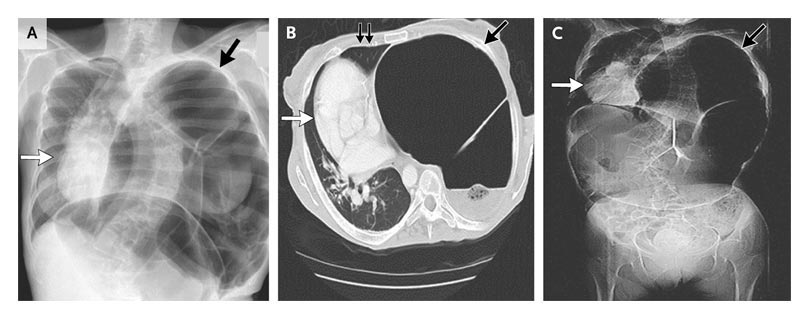
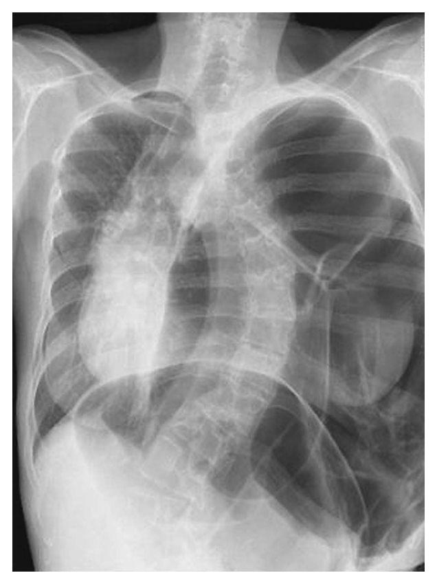
Panel A shows the original chest film, and Panels B and C, both CT scans, show an axial and a scout view of the chest, respectively. Dilated colonic loops occupy the left hemithorax (Panels A, B, and C, single black arrows). The left lung is nearly collapsed (Panel B, double black arrows), with deviation of the mediastinum to the right (Panels A, B, and C, white arrows).
ผู้ป่วยได้รับการตรวจ colonoscopy ซึ่งพบเนื้องอกหนึ่งก้อนที่ transverse colon เธอได้รับการผ่าตัด subtotal colectomy ร่วมกับ ileosigmoid anastomosis จากการผ่าตัด พบว่ามีก้อนขนาดเส้นผ่าศูนย์กลาง 10 ซม.ที่ midtransverse colon และมี eventration ของกระบังลมข้างซ้าย ผลชิ้นเนื้อของก้อนเป็น mucinous adenocarcinoma ลำไส้ใหญ่ขยายตัวอย่างมาก (ขนาดเส้นผ่าศูนย์กลาง 15 ซม.) จาก proximal transverse ไปถึง distal sigmoid portions, โดย มีผนังลำไส้บางมาก (ความหนาโดยเฉลี่ย <0.1 ซม.) แต่ไม่มีไม่มีหลักฐานของการทะลุ พบว่ามี ganglion cells อยู่ในผนังลำไส้ จากการตรวจค้น ไม่พบว่ามี metastasis
ผู้ป่วยได้รับการรักษาด้วย adjuvant chemotherapy และยังไม่พบการกลับเป็นมะเร็งอีกจากการติดตามนาน 15 เดือน
Reference: Karakis I. Medical Mystery: Constipation -- The Answer. NEJM 2009; 360: 1259-60.
Posted by : cpantip , Date : 2010-03-26 , Time : 16:15:05 , From IP : 172.29.3.68
|

