ความคิดเห็นทั้งหมด : 7

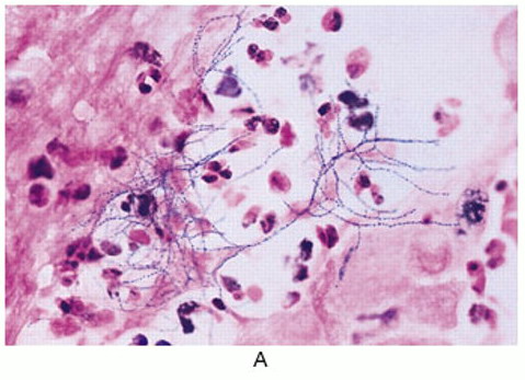
A 20-YOM had fevers with chills, productive cough, and weight loss for 1 month


    ชายอายุ 20 ปี เดิมแข็งแรงดี
-7 เดือนก่อน ผู้ป่วยมีไข้อยู่นาน 3 สัปดาห์จึงไปรพ. การตรวจร่างกาย ตรวจทางห้องปฏิบัติการและภาพรังสีทรวงอกปกติ ต่อมาไข้ลงเอง และผู้ป่วยสบายดี
-1 เดือนก่อน ผู้ป่วยเริ่มมีไข้หนาวสั่น ไอมีเสมหะและน้ำหนักลด ผู้ป่วยได้รับการรักษาที่รพ.แห่งหนึ่งซึ่งแพทย์ให้การวินิจฉัย community-acquired pneumonia และให้การรักษาด้วย ceftriaxone และ azithromycin ต่อมาแพทย์เพิ่มยา clindamycin แต่อาการยังคงเดิม เนื่องจากผู้ป่วยยังมีไข้และไอจึงย้ายผป.มาที่รพ.ใหม่
ผู้ป่วยไม่เคยป่วยเป็นโรคใดๆ ไม่มีประวัติสัมผัสผู้ป่วยโรคใดๆหรือสัตว์ และการเดินทาง ผู้ป่วยเป็นนักศึกษา ไม่สูบบุหรี่ ไม่ดื่มและไม่ติดยาใดๆ
At admission to new hospital, the patient was febrile and dyspneic, with bilateral fine crackles heard on lung examination.
CBC: hemoglobin level, 11 g/dL, WBC, 21,000 cells/μL with a left shift and platelet count, 420,000 platelets/μL). erythrocyte sedimentation rate was 92.
ในรูป A คือภาพรังสีทรวงอก
1. ผู้ป่วยน่าจะเป็นโรคใด
2. จะ manage อย่างไรต่อไป


Posted by : cpantip , E-mail : (chpantip@medicine.psu.ac.th) ,
Date : 2010-03-16 , Time : 15:58:13 , From IP : 172.29.3.68


ความคิดเห็นที่ : 1


   ขอตอบค่ะ
ปัญหาของผป.(ชายอายุ 20 ปี เดิมแข็งแรงดี)
1. ไข้หนาวสั่น ไอมีเสมหะและน้ำหนักลดมา 1 เดือนก่อน ได้รับ Dx CAP รักษาด้วย ceftriaxone + azithromycin + clindamycin แต่อาการยังคงเดิม
2. เคยมีไข้อยู่นาน 3 สัปดาห์ซึ่งหายเองเมื่อ 7 เดือนก่อน
3. CBC: hemoglobin level, 11 g/dL, WBC, 21,000 cells/μL with a left shift and platelet count, 420,000 platelets/μL). erythrocyte sedimentation rate was 92.
4. CXR: necrotizing pneumonia involve the whole right lung
Assessment
1. Necrotizng pneumonia ที่เป็นมานาน 1 เดือน นึกถึง TB, NTM infection, melioidosis, nocardiosis, actinomycosis, fungal infection (histoplasmosis), rhodococcosis ยาที่รักษาอยู่ไม่ครอบคลุมเชื้อเหล่านี้เลย
2. ผู้ป่วยน่าจะมี underlying disease เช่น HIV infection, DM เป็นต้น

Plan: 1. ส่ง anti HIV antibody, FBS และอื่นๆ เพื่อหาว่ามีภาวะภูมิคุ้มกันบกพร่อง
2. ส่ง sputum culture for melioidosis, mycobacteria, fungi, anearobic culture ด้วย และส่งย้อม acid fast stain x 3 วัน, modified acid fast stain , GMS
3. น่าจะทำ bronchoscopy เพื่อเก็บเสมหะมาย้อม และทำ transbronchial biopsy

ยังไม่ให้ antimicrobial agent ใดๆก่อนค่ะ


Posted by : lara , Date : 2010-03-21 , Time : 10:21:01 , From IP : 172.29.3.68

ความคิดเห็นที่ : 2


   คุณหมอ lara ตอบได้ดีมากค่ะ มี differentail diagnosis ครบถ้วน

ผู้ป่วยได้รับการตรวจ chest radiograph (รูป 1) และ CT scan (รูป 2)
การตรวจเสมหะโดย Gram staining, acid-fast bacilli staining, การเพาะเชื้อและ PCR for mycobacterial agents ไม่พบเชื้อใดๆ การเพาะเชื้อของเลือดก็ไม่ขึ้น. biochemical test results และ electrolyte levels (รวม liver function test, lactate dehydrogenase, และ angiotensin-converting enzyme) อยู่ในเกณฑ์ปกติ
Serological tests for HIV and Mycoplasma species, antigen assays for Pneumococus species and Legionella pneumophilia, were all negative. Serum galactomannan assay and PPD test were negative. Antinuclear antibody, anti–double-stranded DNA, and antineutrophil cytoplasm antibody (cytoplasmic and perinuclear) test results were negative.


Posted by : cpantip , Date : 2010-03-21 , Time : 13:20:08 , From IP : 172.29.3.68

ความคิดเห็นที่ : 3


   ผู้ป่วยไม่ตอบสนองต่อการรักษาด้วย vancomycin, ceftazidime, และ clindamycin จึงได้รับการทำ bronchoscopic examination แต่ไม่ได้คำตอบ แพทย์ได้เพิ่มยา isoniazid, rifampin, และ ethambutol ไปนาน 2 สัปดาห์แต่ผู้ป่วยไม่ดีขึ้น สุดท้ายผู้ป่วยได้รับการทำ an open-lung biopsy ซึ่งแสดง necrotizing inflammatory process with granuloma formation, และการเพาะเชื้อของชิ้นเนื้อจากปอดขึ้นเชื้อดังในรูป (ย้อม modified acid-fast bacilli stain)

1. การวินิจฉัยโรคคืออะไร
2. จะ manage อย่างไร


Posted by : cpantip , Date : 2010-03-21 , Time : 13:22:44 , From IP : 172.29.3.68

ความคิดเห็นที่ : 4


   Gram stain ของเนื้อเยื่อจากปอดดังใน slide

Posted by : cpantip , Date : 2010-03-22 , Time : 11:42:05 , From IP : 172.29.3.68

ความคิดเห็นที่ : 5


   จากการย้อม modified acid fast พบเชื้อที่ติดสี acid fast เป็น fillament with branching และมี beading appearance
ส่วนการย้อมสี Gram พบเป็น Gram-positive rod เป็น fillament with branching และมี beading appearance
ตอบ: pulmonary nocardiosis
การรักษา ให้ bactrim -ขนาดของ trimethoprim 15 mkd ไปนาน 2 สัปดาห์แล้วอาจลดขนาดลง เป็น 3 เม็ดเช้าเย็น รักษาไปนาน 6 เดือน


Posted by : lara , Date : 2010-03-25 , Time : 14:35:55 , From IP : 172.29.3.68

ความคิดเห็นที่ : 6


   คุณหมอ lara ตอบถูกต้องแล้วค่ะ
รูป 2. CT of the thorax with contrast demonstrates bilateral consolidation with air bronchograms and cavities
รูป 3+4. Modified acid-fast bacilli stain and gram stain of a culture sample obtained from lung tissue demonstrates partially acid-fast, gram-positive bacteria with characteristic branching and beaded appearance, compatible with Nocardia species.

Diagnosis: severe pulmonary nocardiosis.

หลังเพาะเชื้อของชิ้นเนื้อจากปอด (aerobic incubation) นาน 6 วันก็ขึ้นเป็น rough, yellowish colonies ซึ่งต่อมาพิสูจน์ได้ว่าเป็น Nocardia asteroides

เนื่องจากเป็นการวินิจฉัย nocardiosis ในคนอายุน้อยที่แข็งแรงดีมาก่อน จึงส่งตรวจสำหรับ chronic granulomatous disease (CGD) คือ
1. Nitroblue tetrazolium test (Orpagen Pharma) demonstrated <5% neutrophil color conversion.
2. flow cytometry, which failed to demonstrate any neutrophils with respiratory-burst activity ยืนยันการวินิจฉัย CGD
3. glucose-6-phosphate dehydrogenase level was normal (severe glucose-6-phosphate dehydrogenase deficiency can cause abnormal respiratory burst activity).

การวินิจฉัย: chronic granulomatous disease (CGD) presenting with severe pulmonary nocardiosis.


Posted by : cpantip , Date : 2010-03-26 , Time : 15:32:38 , From IP : 172.29.3.68

ความคิดเห็นที่ : 7


   CGD เป็น phagocytic disorder ซึ่งพบได้น้อย ความผิดปกติที่พบคือ phagocytic cells ไม่สามารถฆ่าเชื้อแบคทีเรียและเชื้อราบางชนิดหลังจากที่กินเชื้อเข้าไป โรคนี้พบในเด็กเป็นส่วนใหญ่ อย่างไรก็ตาม มีรายงานจำนวนหนึ่งที่พบว่าโรคนี้เริ่มปรากฏอาการครั้งแรกในผู้ใหญ่ ความผิดปกติที่จำเพาะของ CGD คือการที่ไม่มี superoxide formation เนื่องจากมีความผิดปกติของ nicotinamide adenine dinucleotide phosphate oxidase enzyme. Enzyme นี้รับผิดชอบในการสร้าง O2− ซึ่งถูกเปลี่ยนไปเป็น bactericidal oxidants ต่างๆ รวมถึง hydrogen peroxide. ทราบกันมานานแล้วว่าโรคนี้มีการถ่ายทอดทางพันธุกรรมแบบ X-linked และ autosomal recessive patterns แต่ specific mutations ที่ทำให้เกิด CGD เพิ่งมีการค้นพบได้ไม่นาน
เนื่องจากคนสามารถใช้ endogenous hydrogen peroxide ซึ่งสร้างขึ้นโดยเชื้อก่อโรค (มากกว่าที่สร้างผ่าน nicotinamide adenine dinucleotide phosphate oxidase enzyme), และเพราะ catalase enzyme ทำให้ hydrogen peroxide สลายไป เชื้อส่วนใหญ่ที่ทำให้เกิดโรคติดเชื้อที่มีลักษณะเฉพาะที่พบใน CGD จึงมี catalase positive ดังนั้น ต้องนึกถึง CGD ในผู้ป่วยที่มีการติดเชื้อที่เกิดจาก catalase-positive organisms ซึ่งอาจเป็นแบบ recurrent หรือที่เกิดขึ้นในตำแหน่งที่พบไม่บ่อย นอกจากนี้ ยังมีการติดเชื้อบางชนิดที่พบได้ยากในคนที่ภูมิต้านทานปกติ ซึ่งต้องหาว่าผู้ป่วยมี underlying CGD ได้แก่ Aspergillus species, Nocardia species, Burkholderia cepacia, Serratia marcescens, Chromobacterium violaceum, Staphylococcus aureus ใน setting ของ liver abscess, และระยะหลังมีรายงาน Granulobacter bethesdensis. ที่น่าสนใจคือ ใน series หนึ่ง หนึ่งในสามของผู้ป่วยที่มี CGD ร่วมกับ nocardiosis มีการติดเชื้อราร่วมด้วย
ผู้ป่วยรายนี้ได้รับการรักษาด้วย IV imipenem และ oral cotrimoxazole ซึ่งทำให้ผู้ป่วยดีขึ้นอย่างรวดเร็ว และผู้ป่วยได้กลับบ้าน 2 สัปดาห์หลังการรักษา หลังรักษาด้วย oral cotrimoxazole นาน 3 เดือน ผู้ป่วยไม่มีอาการใดๆ และรอยโรคในภาพรังสีทรวงอกดีขึ้นอย่างมาก แผนการรักษาคือให้ cotrimoxazole ไปอีก 3 เดือน (รวม 6 เดือน) แล้วตามด้วยการป้องกันด้วยยา cotrimoxazole และ itraconazole. IFN-γ มีประโยชน์ในผู้ป่วย CGD แต่ไม่ได้นำมาใช้กับผู้ป่วยรายนี้เพราะมีราคาแพง

Reference: Heidarnejad H, et al. Acute Necrotizing Pneumonia in a Previously Healthy Young Adult . Clin Infect Dis 2008;46:266–7.


Posted by : cpantip , Date : 2010-03-26 , Time : 15:33:43 , From IP : 172.29.3.68

สงวนสิทธิ์การแสดงความคิดเห็นสำหรับ สมาชิกเท่านั้น