ความคิดเห็นทั้งหมด : 5

A 27-YOM with AIDS had fever and productive cough for 1 weeks


    ผู้ป่วย AIDS เพศชายอายุ 27 ปีมารพ.เนื่องจากมีไข้มา 2 สัปดาห์และไอมีเสมหะมากมา 1 สัปดาห์

1. รูป A คือ CXR ของผู้ป่วย พบความผิดปกติอะไรบ้าง
2. การวินิจฉัยคืออะไร
3. เชื้อก่อโรคน่าจะเป็นเชื้ออะไร
4. จะ manage อย่างไร


Posted by : cpantip , E-mail : (chpantip@medicine.psu.ac.th) ,
Date : 2010-03-05 , Time : 08:37:55 , From IP : 172.29.3.68


ความคิดเห็นที่ : 1


   ขอถามเพิ่มค่ะ
5. ในผู้ป่วยรายนี้ต้องคอยเฝ้าระวังภาวะแทรกซ้อนที่รุนแรงอะไรคะ


Posted by : cpantip , Date : 2010-03-05 , Time : 08:39:26 , From IP : 172.29.3.68

ความคิดเห็นที่ : 2


   ปัญหาของผป.
1. AIDS ไม่ทราบว่า CD4 เท่าไหร่คะ
2. มีไข้มา 2 สัปดาห์และไอมีเสมหะมากมา 1 สัปดาห์
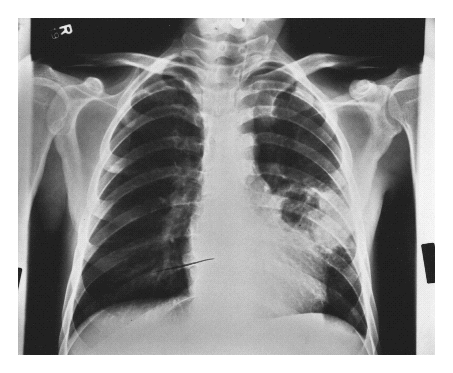
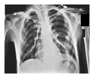
3. CXR: มี lesion ที่ปอดข้างซ้าย ลักษณะกลม มีทั้งลมและส่วนที่ทึบ น่าจะเป็น lung abscess (ประวัติไอมีเสมหะมากด้วย) หรือ necrotizing pneumonia

การวินิจฉัย
1. HIV infection
2. Lung abscess หรือ necrotizing pneumonia เชื้อก่อโรคที่น่าจะเป็น คือ
anaerobe ในปาก, S. aureus, Pneumococci บาง strain, K. pneumoniae
ส่วนเชื้อที่ relate กับ HIV เช่น TB, NTM, nocardia, rhodococcus และ fungi

ก็ต้องเอา sputum มาย้อมสีแกรม, modified acid fast และ acid fast stain
และส่ง culture for aerobic bacteria และ mycobacterium


ต้องคอยเฝ้าระวังภาวะแทรกซ้อนที่รุนแรงอะไร
1. เพราะ abscess อยู่ติดกับหัวใจ ก็ต้องระวังว่าจะแตกเข้า pericardium ได้
2. อาจมี abscess ที่อื่น เช่น nocardia, rhodococcus ก็อาจมี brain abscess ได้ค่ะ


Posted by : lara , Date : 2010-03-12 , Time : 12:40:29 , From IP : 172.29.3.68

ความคิดเห็นที่ : 3


   คุณหมอ lara ตอบได้ถูกต้องแล้วค่ะ

The chest x-ray film in Panel A shows cavitating pneumonia in the left mid-lung field, adjacent to the cardiac silhouette. การเพาะเชื้อของ specimen ที่ได้จาก bronchoscopic sheath catheter ขึ้น Pseudomonas aeruginosa และ Staphylococcus aureus.

ทั้งๆที่ได้ยาปฏิชีวนะที่เหมาะสม ผู้ป่วยมีอาการเจ็บหน้าอกและเหนื่อยซึ่งเกิดขึ้นอย่างรวดเร็ว ตรวจร่างกายพบมี pulsus paradoxus และ JVP สูงถึง mid neck, lungs clear. CXR รูป B พบความผิดปกติอะไรบ้าง
1. การวินิจฉัยคืออะไร
2. จะ manage อย่างไร


Posted by : cpantip , Date : 2010-03-12 , Time : 14:34:46 , From IP : 172.29.3.68

ความคิดเห็นที่ : 4


   ขอลองตอบครับ
-->ข้อ1. CXR รูป B พบความผิดปกติอะไรบ้าง, การวินิจฉัยคืออะไร?
จากรูป B นั้นพบ air density ปริมาณมากใน pericardial sac ทำให้นึกถึงภาะ pneumopericardium ครับ ซึ่งวินิจฉัยได้จากการเห็นลักษณะ air ดังกล่าวร่วมกับอาการปวดหน้าอก (paracardiac radiotransparent band ในผู้ป่วย pleuropericardial-type chest pain) การตรวจพบ JVP สูง(หรือ hypotensionหรือ muffled heart sound) ทำให้สงสัยภาวะ peumopericardium ซึ่งทำให้เกิด cardiac tamponade ซึ่งพบไม่บ่อยมากนัก [โดย intrapericardial pressure ต้องสูงกว่า atrial filling pressure ประมาณ 20-25cmH2O ในคนปกติ]
-->ข้อ2. จะ manage อย่างไร?
แนวทางการรักษาคือ urgent decompression (pericardiocentesis) และติดตาม hemodynamic status ของผู้ป่วยเพื่อยืนยันการวินิจฉัยและติดตามการรักษา สำหรับผู้ป่วยรายนี้พบว่า lung abscess หรือ necrotizing pneumonia ติดกับหัวใจ กล่าวคือมี Silhouette sign คิดว่าอาจเป็นที่มาของ air ใน pericardial sac นี้ได้ ทั้งนี้อาจเกิด fistula ระหว่าง lung parenchyma กับ pericardial sac ดังนั้นในรายนี้คงต้องปรึกษาศัลยแพทย์สำหรับ surgical intervention ปิด fistula ดังกล่าวต่อไป
[ref: Dr. José Barquero Romero, Rev Esp Cardiol. 2005;58(2):227-9
Trujillo MH, Cardiology. 2006;105(1):34-6. Epub 2005 Sep 22.]


Posted by : weeratian , Date : 2010-03-13 , Time : 17:55:14 , From IP : 172.29.22.75

ความคิดเห็นที่ : 5


   คุณหมอ Weeratian ตอบถูกแล้วค่ะ

chest film รูป B: air under pressure is evident in the pericardial sac, with the original abscess cavity now clear of fluid.
ผู้ป่วยมี spontaneous rupture ของฝีในปอดเข้าสู่ pericardial sac และตายจาก tension pneumopericardium และ cardiac tamponade.
รูป C ถ่ายตอน autopsy พบว่า ที่ parietal pericardial surface มี fistula (หัวลูกศร) ซึ่งติดต่อระหว่าง pericardial sac (P) กับ ฝีในปอด

Reference: Smith LG, Naqvi T. Tension Pneumopericardium. NEJM 1995; 332 (22): 1481.


Posted by : cpantip , Date : 2010-03-14 , Time : 16:45:06 , From IP : 172.29.3.68

สงวนสิทธิ์การแสดงความคิดเห็นสำหรับ สมาชิกเท่านั้น