คุณหมอ lara ตอบถูกต้องแล้วค่ะ เก่งมากๆ ค่ะ
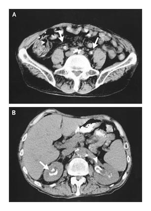
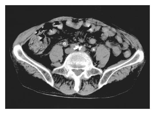
The arrows in Panel A point to the two ureters showing concentric calcification. In Panel B, mural calcification within the left calyx (arrowhead) and the upper pole of the right kidney (arrow) are seen.
ผู้ป่วยมี encrusted pyeloureteritis & cystitis (encrust=ปกคลุมภายนอกด้วยเปลือกแข็ง) ที่เกิดจาก Corynebacterium urealyticum. การส่ง urine culture หลายครั้งขึ้น corynebacterium species ซึ่งต่อมาพิสูจน์ว่าเป็น C. urealyticum. การวินิจฉัยถูกต้องได้มาจากการที่ผล UA หลายครั้งพบว่าผู้ป่วยมี persistently alkaline urine และการที่มี triple phosphate crystals และ concentric calcification ของ ureter ทั้ง 2 ข้างดังที่พบใน computed tomographic scanning ของ pelvis. นอกจากนี้ "contaminants" ในปัสสาวะน่าจะเป็น diphtheroids (common skin flora) แต่จริงๆ แล้วคือ C. urealyticum, ซึ่งแยกได้จากการเพาะเชื้อของปัสสาวะ 10 ครั้งในช่วงเวลา 9 เดือน
ผู้ป่วยได้รับการรักษาด้วยการใส่ nephrostomy tubes ทั้ง 2 ข้าง เพื่อรักษา intermittent urinary obstruction ผู้ป่วยได้รับการรักษาด้วย IV vancomycin นาน 3 สัปดาห์ ซึ่งทำให้ hematuria หายไปและกำจัด C. urealyticum ได้ หน้าที่ของไตดีขึ้นและเขาถูกจำหน่ายไปอยู่ที่ nursing home ต่อมาเขาเสียชีวิตจากภาวะแทรกซ้อนของ aspiration pneumonia.
C. urealyticum เป็น gram-positive, aerobic, urease-positive bacterium with a diphtheroid morphology เชื้อนี้เป็นหนึ่งในจำนวนเชื้อหลายชนิดที่เป็น urea-splitting bacteria (รวมถึง Proteus mirabilis), ซึ่งสามารถเปลี่ยน urea เป็น ammonia ทำให้เกิดปัสสาวะที่เป็นด่าง กระบวนการนี้สามารถมีผลทำให้เกิดการตกตะกอนของ struvite และ calcium phosphate crystals ซึ่งทำให้เกิด kidney stones (รูป A) และ encrustation ทั่วทางเดินปัสสาวะ (รูป B). Pyelitis และ cystitis เป็นผลจากการอักเสบที่เรื้อรัง
Reference: Yu V L, Kim DHD. Medical mystery: concentric calcification--the answer.. NEJM 2006;354: 1433-1434.
Posted by : chpantip , Date : 2010-01-08 , Time : 10:23:13 , From IP : 172.29.3.68
|