คุณหมอ sunshine ตอบได้ถูกต้องแล้วค่ะ
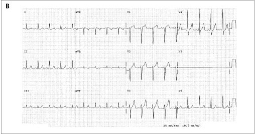
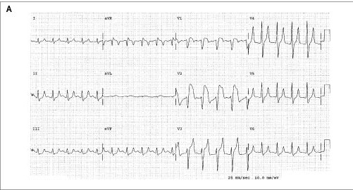
The initial electrocardiogram (รูป A) revealed sinus tachycardia and ST-segment elevation in leads V1 to V3 findings highly suggestive of acute anteroseptal myocardial infarction. Peaked T waves were noted in leads II, III, aVF, and V3 to V6.
ได้ส่ง serum glucose concentration = 839 mg/dL, arterial blood pH = 7.21, และ serum potassium concentration = 7.9 mmol per liter.
การวินิจฉัยคือ diabetic ketoacidosis
ได้ทำ electrocardiogram ซ้ำหลายชั่วโมงต่อมาเมื่อ potassium concentration ลดลงเป็น 5.1 mmol per literจากการรักษา (รูป B [ไม่ได้ใส่รูป lead V5 ]) ST-segment elevation disappeared completely, as did the peaked T waves.
ผู้ป่วยรายนี้เป็นตัวอย่างของ hyperkalemia ที่ทำให้เกิด "pseudoinfarction" pattern. สิ่งสำคัญสำหรับการวินิจฉัยที่ถูกต้องคือ T wave ใน V4, (which is tall, narrow, and pointed), with a short QT interval. Tall T waves ซึ่งเป็นลักษณะที่จำเพาะของ hyperacute ischemic changes มักจะสัมพันธ์กับ long QT interval, และ the T waves are broad rather than narrow and pointed.
Reference: Wang K. Pseudoinfarction pattern due to hyperkalemia. NEJM 2004; 351 (6): 593.
Posted by : chpantip , Date : 2009-12-21 , Time : 11:07:10 , From IP : 172.29.3.68
|

