ความคิดเห็นทั้งหมด : 4

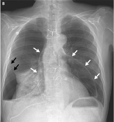
A 60-YOM had progressive dyspnea 3 mo. after AVR +mitral valve repair


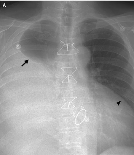
   ชายอายุ 60 ปี มีอาการหอบเหนื่อยมานาน 3 เดือนหลังจากได้รับการผ่าตัด aortic-valve replacement และ mitral-valve repair สำหรับ valvular insufficiency ที่สัมพันธ์กับการเป็น infective endocarditis มาก่อน หลังการผ่าตัดไม่มีภาวะแทรกซ้อนใดๆ .

1. ในรูป A คือ ภาพรังสีทรวงอกเมื่อแรกรับ พบความผิดปกติอะไรบ้าง
2. การวินิจฉัย น่าจะเป็นอะไร
3. จะ manage อย่างไร


Posted by : chpantip , E-mail : (chpantip@medicine.psu.ac.th) ,
Date : 2009-12-17 , Time : 10:09:06 , From IP : 172.29.3.68


ความคิดเห็นที่ : 1


   ขอลองตอบครับ
1. ลูกศรคือ Right pleural effusion หัวลูกศรคิดว่าเป็น double shadow sign >> pericardial effusion
2. คิดว่าจากประวัติเหนื่อยหลังจาก cardiac surgery ที่มี pericardiotomy ในช่วงแรก Film พบ effusion >> คิดว่าเป็น postpericardiotomy syndrome
3. ตรวจ sign of cardiac temponade, Echocardiogram + Drainage ที่ pleural effusion +/- pericardial effusion >> ให้ antiinflam เช่น NSAID, Steroid ส่วน colchicine ไม่แน่ใจว่า prove แล้วหรือไม่


Posted by : sunshine , Date : 2009-12-20 , Time : 11:54:34 , From IP : 172.29.22.198

ความคิดเห็นที่ : 2


   ยินดีต้อนรับ คุณหมอ sunshine คุณหมอตอบได้ดีค่ะ

A chest radiograph obtained on presentation (รูป A) revealed a large pleural effusion on the right side (arrow) and an increase in the cardiac silhouette (arrowhead). Computed tomography confirmed the presence of the pleural effusion and revealed a moderate pericardial effusion.
ผู้ป่วยได้รับการใส่ pigtail drainage catheter เข้าไปใน pleural cavityและ exudative pleural fluid 2.8 liters ถูกระบายออกมา.

1. รูป B คือภาพรังสีทรวงอกที่ทำหลังการทำหัตถการ พบความผิดปกติอะไรบ้าง
2. จะ manage อย่างไรต่อไป


Posted by : chpantip , Date : 2009-12-21 , Time : 11:01:13 , From IP : 172.29.3.68

ความคิดเห็นที่ : 3


   1 รูป B ลูกศรสีดำคือ pneumothorax ครับ
ลูกศรสีขาวคือ pneumopericardium ครับ ซึ่งต้องแยกจาก pneumomediastinum ครับ
2. ใส่ chest drian + pericardial drian and F/U CXR ครับ


Posted by : sunshine , Date : 2009-12-22 , Time : 19:19:29 , From IP : 172.29.22.198

ความคิดเห็นที่ : 4


   A chest radiograph obtained after the procedure (รูป B) showed a large pneumothorax on the right side (black arrows), with associated atelectasis in the lower lobe, as well as a large pneumopericardium (white arrows).

ได้คา percutaneous drainage ไว้ต่อไป และต่อมา ผู้ป่วยได้รับการรักษาด้วย antiinflammatory drugs และ colchicine สำหรับ postpericardiotomy syndrome ผู้ป่วยมีอาการดีขึ้นตามลำดับและเป็นปกติดีเมื่อกลับบ้าน และสบายดีหลังจากนั้น


Reference: Andrade J, Aymong E. Pneumopericardium and pneumothorax. NEJM 2009; 361 (5): 510.


Posted by : chpantip , Date : 2010-01-01 , Time : 16:05:36 , From IP : 172.29.3.68

สงวนสิทธิ์การแสดงความคิดเห็นสำหรับ สมาชิกเท่านั้น