ความคิดเห็นทั้งหมด : 2

An 83-YOW was hospitalized with nausea, vomiting, and obstipation


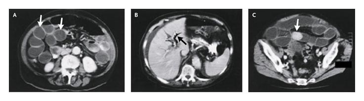
   หญิงอายุ 83 ปีเข้ารับการรักษาในรพ.เนื่องจากคลื่นไส้ อาเจียนและไม่ถ่ายอุจจาระ
ผู้ป่วยมีน้ำหนักลดมานาน 6 เดือนและมีคลื่นไส้ อาเจียนและท้องผูกซึ่งเป็นไม่รุนแรงในช่วงแรก อาการเป็นมากขึ้นเรื่อยๆ
เธอไม่เคยได้รับการผ่าตัดในช่องท้องและไม่มีโรคใดๆมาก่อน
The physical examination revealed a distended abdomen, with no palpable masses and no hernias.

เพื่อแยก ileus จาก mechanical small-bowel obstruction, ผู้ป่วยจึงได้รับการตรวจ enhanced computed tomography of the abdomen and pelvis.
1. จาก CT นี้ พบความผิดปกติอะไรบ้าง
2. การวินิจฉัยคือ................................................
3. จะ manage อย่างไร


Posted by : chpantip , E-mail : (chpantip@medicine.psu.ac.th) ,
Date : 2009-12-02 , Time : 13:44:58 , From IP : 172.29.3.68


ความคิดเห็นที่ : 1


   ขอตอบค่ะ
ผู้ป่วยมี intestinal obstruction vs ileus
จาก CT abdomen พบว่ามีลำไส้ dilate, pneumobilia และมีก้อนทึบอยู่ในลำไส้
เข้าได้กับ Rigler triad (The classic roentgenographic signs described by Rigler et al, include intestinal obstruction, pneumobilia, aberrantly located gallstone and change of location of the previously identified stone on serial exams) ค่ะ

การวินิจฉัยคือ gallstone ileus

การรักษา ผ่าตัดเอาก้อนนิ่วที่อุดตันลำไส้ออก


Posted by : lara , Date : 2009-12-08 , Time : 14:13:24 , From IP : 172.29.3.68

ความคิดเห็นที่ : 2


   คุณหมอ lara ตอบถูกแล้วค่ะ

enhanced computed tomography of the abdomen and pelvis: numerous distended loops of small intestine (รูป A, ลูกศร), air in the biliary tree (รูป B, ลูกศร), and a calcified intraluminal mass (รูป C, ลูกศร) were identified.

การตรวจพบทั้งหมดทำให้สามารถวินิจฉัย gallstone ileus
ผู้ป่วยได้รับการผ่าตัด laparotomy with enterolithotomy และไม่ทำอะไรกับ gallbladder
ผู้ป่วยหายดีหลังจากนั้น

Reference: Graham JS, Rothwell BC. Gallstone Ileus. NEJM 2004; 351 (11): 1119.


Posted by : chpantip , Date : 2009-12-10 , Time : 15:38:59 , From IP : 172.29.3.68

สงวนสิทธิ์การแสดงความคิดเห็นสำหรับ สมาชิกเท่านั้น