ความคิดเห็นทั้งหมด : 2

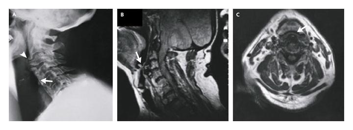
An 81-YOM had increasing shortness of breath and throat tightness,


   ชายอายุ 81 ปีมีอาการกลืนลำบากเล็กน้อยมาหลายปี ในช่วง 6 เดือนก่อนเข้ารักษาในรพ. อาการกลืนลำบากเป็นมากขึ้น และมีไอเป็นเลือดเป็นครั้งคราว
หลายวันก่อนเข้ารับการรักษาในรพ. ผู้ป่วยเหนื่อยง่ายและแน่นในคอ อาการทั้ง 2 อย่างเป็นมากขึ้นในท่านอนหงาย
Laryngoscopic examination revealed marked narrowing of the airway due to a visible retropharyngeal bulge.

ตรวจร่างกายพบว่ามี stridor จึงรับผู้ป่วยใน ICU
ผู้ป่วยได้รับการตรวจ radiographs of the cervical spine และ magnetic resonance images (MRI) of the neck ทันที
รูป A คือ lateral radiograph of the cervical spine
รูป B คือ sagittal MRI scan
รูป C คือ axial MRI scan
1. พบความผิดปกติอะไรบ้าง
2. การวินิจฉัยคือ............................................
3. จะ manage อย่างไร


Posted by : chpantip , E-mail : (chpantip@medicine.psu.ac.th) ,
Date : 2009-11-25 , Time : 16:00:03 , From IP : 172.29.3.68


ความคิดเห็นที่ : 1


   จากรูป C-spine lateral ที่ anteroir part มี bone หนาๆ แปลกๆ ยื่นออกมา ตั้งแต่ C2-6 น่าจะเป็นสาเหตุทำให้มี airway obstruction คิดว่าเป็น bone spur
คงต้องถามคน intubation ว่าใส่ ETT ลำบากหรือเปล่า
management คงต้องพึ่ง surgeon remove bone ออกน่าจะ definite ที่สุด


Posted by : blackbear , Date : 2009-11-29 , Time : 14:24:16 , From IP : 172.29.3.84

ความคิดเห็นที่ : 2


   คุณหมอ blackbear ตอบถูกแล้วค่ะ เก่งมากๆ ค่ะ เพราะ film อ่านยาก ขอลงรูปให้ได้อ่านประกอบกับผลการอ่านฟิล์มนะคะ

The lateral radiograph of the cervical spine revealed a huge mantle of osteophytic bone anterior to the spine from C2 to C7, with fusion of the osteophytes from C4 to C7 (iรูป A, arrow). Osteophytes at C2 and C3 compressed the hypopharyngeal airway at the level of the epiglottis (รูป A, arrowhead). A sagittal MRI scan showed anterior displacement of the prevertebral soft tissues by these osteophytes (รูป B, arrow), and an axial scan showed compression of the airway to a slit-like opening (รูป C, arrow).
หลังจากการใส่ endotracheal tube ซึ่งใส่ยากมาก neurosurgeon และ otolaryngologist ได้ทำ anterior resection of the ventral spinal osteophytes with partial diskectomies at C2–C3 and C3–C4.
ผู้ป่วยดีขึ้นหลังการผ่าตัดและหายเป็นปกติจากการติดตามที่ 1 เดือนหลังจากกลับบ้าน

Reference: Aronowitz P, Cobarrubias F. NEJM 2003; 349: 2540.


Posted by : chpantip , Date : 2009-12-02 , Time : 14:22:24 , From IP : 172.29.3.68

สงวนสิทธิ์การแสดงความคิดเห็นสำหรับ สมาชิกเท่านั้น