ความคิดเห็นทั้งหมด : 5

A 52-YOM had right-sided pleurisy, fever, and shaking chills for 3 days.


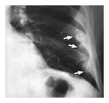
   ชายอายุ 52 ปีมารพ.ด้วยอาการเจ็บหน้าอกข้างขวาโดยเฉพาะเวลาไอหรือหายใจลึกๆ มีไข้และหนาวสั่น เป็นมา 3 วัน.

On examination he was febrile and had marked tenderness to percussion over the right lower rib cage.

ในภาพรังสีทรวงอกของผป.พบว่ามี elevated right hemidiaphragm
1. รูป A คือ left lower lung field พบความผิดปกติอะไรบ้าง
2. จะ manage อย่างไรต่อไป


Posted by : chpantip , E-mail : (chpantip@medicine.psu.ac.th) ,
Date : 2009-10-05 , Time : 10:37:30 , From IP : 172.29.3.68


ความคิดเห็นที่ : 1


   Psittacosis
1. CXR เห็นรูป decrease lung volume >>wedge shape , fracture of anterior part of left ribs
2. Management
Tetracyclines 2 g daily แบ่งให้ 4 dose 7-14 วัน
หรือ Doxycycline (100 mg by mouth bid)
ในกรณีแพ้ยากลุ่มข้างต้นอาจพิจารณาใช้ Erythromycin


Posted by : LILO , Date : 2009-10-05 , Time : 20:16:31 , From IP : 118.173.1.67.adsl.dy

ความคิดเห็นที่ : 2


   case นี้มาด้วยอาการไข้หนาวสั่น คิดถึงโรคในกลุ่ม acute febril illness: malaria และ bacteremia อาการ pleurisy และตรวจร่างกาย marked tenderness to percussion over the right lower rib cage ไม่อธิบาย malaria

source of infection
1. Respiratory system
--> parenchymal disease : right lower lobe pneumonia
-->pleural disease : emphyema, pleural effusion
2. Hepatobilliary tract infection ที่มี diaphragmatic irritation
--> liver abscess
-->chplecystitis

จาก CXR. พบว่ามี elevated right hemidiaphragm
ภาพ left lower lung field : multifocal poorly marginated peripheral lung nodules +/- cavity

รายนี้มีปัญหา fever with chill with tender hepatomegaly CXR ไม่มี lung infiltration ชัดเจนที่จะอธิบาย pneumania ทำให้คิดถึง liver abscess มากที่สุด

ภาพในข้อ 1. คิดว่าเข้าได้กับ septic pulmonary embolism เพราะ
(1) multifocal lung infiltrates
(2) presence of active extrapulmonary infection as potential
embolic source --> liver abscess
(3) exclusion of other potential explanation for lung infiltrates

manage :
1.investigation : ulstrasound ABD CT with contrast evaluate lesion and organ involvement
2.Get rid of infection : ATB, hepatic drainage


Posted by : tiplada , Date : 2009-10-05 , Time : 20:33:32 , From IP : 118.173.129.81.adsl.

ความคิดเห็นที่ : 3


   คุณหมอ tiplada ตอบถูกแล้วค่ะ

chest radiograph showed an elevated right hemidiaphragm and several lesions characteristic of septic pulmonary emboli in the left lower lobe (รูป A, arrows).
Abdominal ultrasonography demonstrated a large, complex, predominantly hypoechoic mass in the posterior segment of the right hepatic lobe (รูป B, arrows).


Posted by : chpantip , Date : 2010-01-03 , Time : 17:06:17 , From IP : 172.29.3.68

ความคิดเห็นที่ : 4


   Contrast-enhanced abdominal computed tomography showed involvement of both the hepatic parenchyma (รูป C, arrows) and the right hepatic vein (รูป D, arrows).

Transthoracic echocardiogram was normal.
Aspiration of the hepatic lesion yielded thick pus. Cultures of the pus and blood grew Streptococcus milleri.

หลังการรักษาด้วย ceftriaxone IV (นาน 2 สัปดาห์ต่อมาเปลี่ยนเป็นกิน penicillin V) และ percutaneous drainage ของฝีในตับ รอยโรคที่ปอดและที่ตับค่อยๆหายไป
S. milleri เป็นส่วนหนึ่งของ flora ของ mucous membranes ในผป.รายนี้ยังไม่มีหลักฐานของตำแหน่งการติดเชื้อที่เข้าสู่กระแสเลือด

Reference: Fred H, Basaria L. Liver Abscess with Septic Pulmonary Emboli.
NEJM 1997;336 (20): 1428.


Posted by : chpantip , Date : 2010-01-03 , Time : 17:07:24 , From IP : 172.29.3.68

ความคิดเห็นที่ : 5


   Contrast-enhanced abdominal computed tomography showed involvement of both the hepatic parenchyma (รูป C, arrows) and the right hepatic vein (รูป D, arrows).


Posted by : chpantip , Date : 2010-01-03 , Time : 17:07:51 , From IP : 172.29.3.68

สงวนสิทธิ์การแสดงความคิดเห็นสำหรับ สมาชิกเท่านั้น