ความคิดเห็นทั้งหมด : 3

A 44-YOW had intermittent discomfort in right upper abdomen


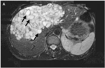
   หญิงอายุ 44 ปีมารพ.เนื่องจากมีอาการแน่น ไม่สบายที่ท้องส่วนบนข้างขวามานาน 10 เดือน
On physical examination, a mass filling the epigastrium and the right subcostal region was noted.

Ultrasonography and computed tomography of the abdomen showed multiple cysts in both lobes of the liver.

An image of the liver obtained by magnetic resonance imaging showed a cystic mass involving the medial segment of the left lobe and the anterior segment of the right lobe (รูป A, arrows).

1. การวินิจฉัยคือ...................................
2. จะ manage อย่างไรบ้าง


Posted by : chpantip , E-mail : (chpantip@medicine.psu.ac.th) ,
Date : 2009-08-31 , Time : 09:50:29 , From IP : 172.29.3.68


ความคิดเห็นที่ : 1


   ขอลองตอบครับ
-->1. การวินิจฉัยคือ........
จากอาการไข้และปวดท้อง(right upper quadrant) มี differential diagnosis มากมาย ได้แก่ โรคปอด pneumonia โดยเฉพาะที่ basal segment, โรคทาง GI ได้แก่ gastritis หรืออาจเป็น โรคตับและท่อน้ำดี ได้แก่ cholecystitis, liver abscess หรือ hydatid cysts โดยทางภาพ ultrasound ดังภาพนั้นก็ช่วยแยกโรคได้ ซึ่งลักษณะที่เห็นคือ multiple cysts ทำให้สิ่งที่นึกถึงได้แก่ polycystic liver disease (PCLD), parasitic or hydatid (echinococcal) cysts, cystic tumors และ abscesses โดยทั่วไปก็แยกกันยาก
-->2. จะ manage อย่างไรบ้าง
1. ส่ง tumor marker ซึ่ง primary tumor ที่พบบ่อยได้แก่ colon, pancreas และ breast
2. parasitologic serologic test (เช่น enzyme-linked immunosorbent assay) เนื่องจากการเจาะมาตรวจอ่านต้องเสี่ยงกับ anaphylactic reaction จาก protoscolices แต่ก็มีวิธีการป้องกันคือการให้ antihistamine และ aspiration แบบ PAIR(Puncture, Aspiration, Injection of an heminthicide and Reaspiration)
[Ref: Mike Stocksley, Abdominal Ultrasound p42-p50]


Posted by : weeratian , Date : 2009-09-01 , Time : 09:58:21 , From IP : 172.29.18.149

ความคิดเห็นที่ : 2


   คุณหมอ Weeratian ตอบถูกแล้วค่ะ

Serologic testing for hydatid disease (enzyme-linked immunosorbent assay) และ complement-fixation test ได้ผลบวก (titer, 1:8).

ผู้ป่วยได้รับการผ่าตัด radical excision ของ cyst (รูป B) และ microscopical examination ของ specimen ยืนยันการวินิจฉัย alveolar hydatid disease. ผู้ป่วยได้รับการรักษาด้วย albendazole
หลังการผ่าตัด เธอหายดี

ขอถามต่อค่ะ ผู้ป่วยเป็นโรค hydatid disease ได้อย่างไร


Posted by : chpantip , Date : 2009-09-04 , Time : 15:42:15 , From IP : 172.29.3.68

ความคิดเห็นที่ : 3


   ผู้ป่วยเป็นโรค hydatid disease ได้อย่างไร
Hepatic alveolar echinococcosis เป็นโรคปาราสิตที่พบไม่บ่อยนัก สาเหตุเกิดจาก E. multilocularis ซึ่งมี endemic area อยู่ที่ central และ northern Europe, northern Asia, Japan, China, Turkey และบางบริเวณของ North America
การเจริญเติบโตนั้นต้องปกติต้องอาศัย sylvatic cycle ใน wild canid ซึ่งเป็น definitive host และสัตว์กัดแทะหลายชนิดซึ่งเป็น intermediate host ตัวเต็มวัยอาศัยอยู่ใน proximal small intestine ของ definitive host โดยเฉพาะอย่างยิ่ง red fox ส่วน intermediate host ติดโดยการกิน wild berry หรือ น้ำ ที่มีไข่ของปาราสิตซึ่งอยู่ในอุจาระของหมาป่าที่ติดเชื้อ น้ำย่อยจาก host เองจะทำลายผนังไข่จึงทำให้ embryo เจาะทะลุ intestinal wall ของ host และเข้าไปถึงตับโดยทาง portal หรือ lymphatic system ที่ซึ่ง larva เจริญเติบโตอย่างมาก
ส่วน intermediate host ที่ไม่อยู่วงจรทั่วไป ได้แก่ มนุษย์ จะเกิดพยาธิสภาพเป็นก้อนโตช้า ๆ ในตับแต่ life-threatening จึงทำให้การวินิจฉัยล่าช้าหลายปี
[Ref: Proietti S, Radiographics. 2004 May-Jun;24(3):861-5.]


Posted by : weeratian , Date : 2009-09-06 , Time : 20:05:23 , From IP : 113.53.2.208

สงวนสิทธิ์การแสดงความคิดเห็นสำหรับ สมาชิกเท่านั้น