ขอลองตอบครับ
ขอลองวิเคราะห์เกี่ยวกับ Chest X-ray ครับ: Chest radiography revealed bilateral multinodular pneumonia and pleural effusions การที่พบว่ามี multiple nodules นั้นช่วยในการวินิจฉัยได้มาก เพราะมีเพียง 5 กลุ่มโรคที่เรานึกถึงซึ่งประกอบด้วย 1.lung metastasis (ภาพที่อยากเห็นคือเป็น หลาย nodule ขนาดไม่เท่ากัน, เด่น lower lobe โดยเฉพาะบริเวณชายปอดเพราะมาจาก hematogenous, bilateral, nodule กลม) 2.Infection ได้แก่ Granulomatous เช่น TB, Nocardiosis, Histoplasmosis etc. และ Collagen vascular disease เช่น rheumatoid nodule และ Wegener’s granulomatosis 3.Septic embolism (มีลักษณะคล้ายกับ lung metastasis แต่ขอบเขตของ nodule จะไม่ค่อยชัด มีการเปลี่ยนแปลงรวดเร็ว) 4.primary tumor ชนิด adenocarcinoma โดยเฉพาะ Bronchioloalveolar carcinoma ซึ่งไม่สัมพันธ์กับการสูบบุหรี่ 5.Pulmonary AVM มักไม่มีอาการแต่หากมีอาการจะเป็นลักษณะ Right to left shunt หรือ brain abscess (Ref: Chest X-ray อ.วิวัฒนา ถนอมเกียรติ) จึงอยากเห็นภาพ CXR ในผู้ป่วยรายนี้มาก อยากไรก็ตามสิ่งคิดถึงหรือเข้าได้กับผู้ป่วยรายนี้มากก็อาจะเป็น active TB เนื่องจากมีอาการไข้ เหนื่อยง่าย อ่อนเพลีย น้ำหนักลด ในช่วงระยะเวลา 2 สัปดาห์ อีกทั้งยังมีหลักฐานทาง CXR คือ pleural effusion ซึ่งเป็น indication ตัวหนึ่งซึ่งบอกว่าเป็นลักษณะที่ active
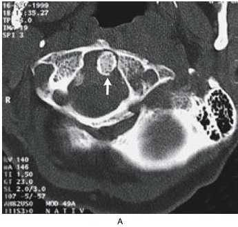
-->1. ในรูป A คือ axial sections ของ computed tomographic scan ของคอพบความผิดปกติอะไรบ้าง
เป็น film CT ตำแหนง upper cervical spine (C1+C2) พบ low attenuation with rim enhancement ด้านซ้ายหลังต่อ cervical spine คิดว่าน่าจะเป็น abscess ของบริเวณ deep neck space ซึ่งตรงนี้น่าจะอธิบายเรื่องกลืนลำบากของผู้ป่วยได้ นอกจากนี้ที่ลูกศรชี้คือ dens ของ C2(axis) ซึ่งเลื่อนไปทางด้านซ้าย ทำให้ LADI ของทั้งสองข้างไม่เท่ากัน อาจเรียกภาวะนี้ว่า atlanto-axial subluxation
-->2. การวินิจฉัยที่น่าจะเป็นมากที่สุดคือ.......
Deep neck abscess หากที่ปอดและบริเวณคอเป็นโรคเดียวกันก็นึกถึง tuberculous osteomyelitis ด้วย
-->3. จะ manage อย่างไรต่อไป
1.ที่ปอด คงต้องเก็บ sputum มาตรวจ: G/S และ AFB, culture
2.ที่ spine คงต้องอาศัย surgical drainage และ broad-spectrum antibiotics สำหรับกรณีที่ต้องทำ emergency drainage คือมี neurologic deficit หลังจากนั้นก็นำ specimen มาตรวจ G/M, AFB และ culture
3.ตรวจร่างกายเพิ่มเติมและเฝ้าระวัง สิ่งที่อันตรายใน deep neck infection ในบริเวณนี้คือการแพร่กระจายไปยัง mediastinum ซึ่งอาจทำให้เกิด pericarditis ได้
Posted by : weeratian , Date : 2009-08-21 , Time : 11:27:42 , From IP : 172.29.13.140
|