ความคิดเห็นทั้งหมด : 5

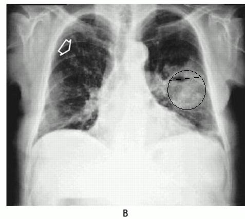
A 76-YOM with Churg–Strauss syndrome had painful "bumps" on his scalp


   ชายอายุ 76 ปีมารพ.เนื่องจากเบื่ออาหาร อ่อนเพลียและเจ็บที่ก้อนที่หนังศีรษะซึ่งมีอยู่หลายก้อน ก้อนบางก้อนแตกออกมาเป็นหนองสีครีม เขามีโรคเดิมคือ Churg–Strauss syndrome ซึ่งได้รับการรักษาด้วย prednisolone ขนาดสูง และ methotrexate อยู่

Examination revealed several partially crusted furuncular lesions as well as enlargement of occipital and cervical lymph nodes.

ครั้งแรก ผู้ป่วยได้รับการรักษาด้วย dicloxacillin และต่อมาแพทย์ได้เปลี่ยนเป็น azithromycin อาการของผู้ป่วยไม่ดีขึ้นเลย ต่อมาผู้ป่วยมีไข้และหนาวสั่น
เขาไม่มีอาการปวดหัว เจ็บหน้าอก หรือไอเลย

Reexamination revealed a 3-cm erythematous, nonfluctuant soft-tissue mass on the back in addition to the scalp lesions and adenopathy. There was no clinical evidence of bone, joint, or central nervous system involvement.

จากการเจาะที่ก้อนที่หนังศีรษะ ได้หนองสีครีม
1. ในรูป A คือ สเมียร์ของหนองย้อมสีแกรม พบความผิดปกติอะไรบ้าง
2. การวินิจฉัยคือ.......................................................
3. จะmanage อย่างไร


Posted by : chpantip , E-mail : (chpantip@medicine.psu.ac.th) ,
Date : 2009-08-10 , Time : 11:00:15 , From IP : 172.29.3.68


ความคิดเห็นที่ : 1


   ขอตอบครับ
-->ข้อ 1. ในรูป A คือ สเมียร์ของหนองย้อมสีแกรม พบความผิดปกติอะไรบ้าง
Branching, beaded, filamentous bacteria จะเห็นว่าติดสีย้อมด้วย Gram-stain ไม่ดีเนื่องจากไม่ถูก decolorized ด้วย acid alcohol หรือเรียกได้ว่ามีคุณสมบัติ acid fastness
-->ข้อ 2. การวินิจฉัยคือ............
ลักษณะของ Branching, beaded, filamentous bacteria เป็นลักษณะของ Nocardia ครับ ซึ่งปัจจัยเสียงในผู้ป่วยรายนี้คือ Immunocompromise เนื่องจากการกินยา prednisolone ขนาดสูง และ methotrexate แม้ว่า Pulmonary disease จะเป็น predominant clinical presentation (more than 40% of reported cases) แต่ยังมี presentation ในรูปแบบของ disseminated infection ได้ เช่น CNS, eye โดยเฉพาะอย่างยิ่ง retina, skin, และ subcutaneous tissues, kidney, joint, bone และ หัวใจ ซึ่งในผู้ป่วยรายนี้พบ skin lesion เป็นอาการแสดงหลัก
จึงขอตอบว่าเป็น disseminated nocardial infection
-->ข้อ 3. จะmanage อย่างไร
1.ต้องหา primary source ซึ่งบริเวณที่พบบ่อยคือ lung คิดว่าควรส่ง Chest X-ray ซึ่งพยาธิสภาพที่เห็นจะเป็น irregular nodules (ซึ่งมักจะเป็น cavitating when large), reticulonodular or diffuse pneumonic infiltrates, and pleural effusions หากไม่สามารถอธิบายว่า lung เป็น primary ได้ ก็อาจต้องไปหาที่อื่นเช่น Brain เพราะว่าพบบ่อยเช่นกัน (44% of cases of all systemic nocardiosis) พยาธิสภาพมักจะเป็น granulomata or abscesses in the brain
2.นำหนองที่เจาะได้ไป culture และทำ blood culture (เพราะว่าเป็น disseminated disease)
3.การรักษานั้นประกอบด้วย surgical drainage or debridement และการให้ยา antibiotics ซึ่งยาที่ใช้คือ กลุ่ม Sulfonamides ใน dose ที่สูงกว่าปกติ (15 mg/kg TMP and 75 mg/kg SMX) เนี่องจากเป็น disseminated disease เริ่มแรกใช้เป็น IV และเปลี่ยนเป็น oral ได้เมื่อ 3-6 สัปดาห์ ซึ่งเป็น HIV-negative immunosuppressed patient การรักษาจะใช้ระยะเวลา 12 months หรือนานกว่านี้
Ref: TANIA C. SORRELL, Chapter 252 – Nocardia Species, Mandell, Bennett, & Dolin: Principles and Practice of Infectious Diseases, 6th ed.


Posted by : weeratian , E-mail : (weeratian@hotmail.com) ,
Date : 2009-08-10 , Time : 18:45:22 , From IP : 172.29.11.74


ความคิดเห็นที่ : 2


   คุณหมอ Weeratian ตอบถูกแล้วค่ะ

Aspiration of a scalp lesion produced creamy purulent material, which contained polymorphonuclear leukocytes and numerous beaded, branching, filamentous bacilli on Gram staining (Panel A).

การเพาะเชื้อของหนองและเลือดขึ้น Nocardia farcinica

รูป B คือ chest radiograph ของผู้ป่วย พบความผิดปกติอะไรบ้าง


Posted by : chpantip , Date : 2009-08-12 , Time : 11:44:20 , From IP : 172.29.3.68

ความคิดเห็นที่ : 3


   ขอลองอ่าน Chest X-ray ครับ
ผู้ป่วยชายไทยอายุ 76 ปี
1.position: AP supine, poor inspiration
2.exposure: ไม่ดีมาก เพราะมองเป็นเห็น upper lung field ไม่หมด
3.Trachea shift to the right อาจเนื่องจาก prominent aorta ที่พบได้ปกติในผู้ป่วยอายุมาก
4.Heart: ขนาดบอกยากในท่า AP, ขอบเรียบและที่ apex พบว่าขอบไม่เรียบตรงนี้จะเป็นเป็น normal variance ซึ่งเรียกว่า epicardial fat pad (ซึ่ง fat ตรงนี้อาจมีประโยชน์ในการวินิจฉัย pericardial effusion)
5.Lung: Diffuse nodular infiltration in Right lung และเด่นที่ upper lung field ตามที่ลูกศรชี้ ส่วน left lung เห็น thick-walled cavity ขนาดประมาณ 5x5cm ซึ่งมี air-fluid level ซึ่งอาจเกิดจากการ aspiration หรือ bronchopleural fistula, น่าจะมี atelectesis บริเวณ Rt. middle lobe ในระดับ segmental หรือ subsegmental เนื่องจากพบ Silhouette sign กับ heart ด้านขวาด้วย
6.Hidden area: โดยทั่วไปก็ปกติดี แต่พบ small nodules ประมาณ 3-4 nodule หลัง cardiac shadow บริเวณ left lower lung field
7.Hilum: ปกติ เนื่องจากระดับด้านซ้ายก็สูงกว่าด้านขวาได้นิดหน่อย, รูปร่างยังไม่กลมหรือผิดรูปไปมาก, ขนาดยังไม่โต, และ density ก็ยังไม่เด่นกว่า cardiac shadow
8.Bone ไม่พบ fracture


Posted by : weeratian , E-mail : (weeratian@hotmail.com) ,
Date : 2009-08-12 , Time : 15:45:22 , From IP : 172.29.11.203


ความคิดเห็นที่ : 4


   สำหรับ thick-walled cavity (ขอยืมรูปอาจารย์ครับ)
มี differential diagnosis ได้ดังนี้
1.lung abscess, necrotic lung cancer, necrotizing pneumonia
2.TB, melioidosis, nocardia, rhodococcus
3.Inflammation: progressive massive fibrosis
4.diaphragmatic hernia (gastric/duodenum)
5.loculated empyema
ref Chest X-RAY - อ.วิวัฒนา ถนอมเกียรติ


Posted by : weeratian , Date : 2009-08-12 , Time : 16:10:33 , From IP : 172.29.11.203

ความคิดเห็นที่ : 5


   คุณหมอ Weeratian ตอบถูกต้องแล้วค่ะ

Chest radiograph disclosed a 7-cm cavitary lesion with an air–fluid level in the left upper lobe and small nodular densities (arrow) in the right upper lung field.

ผู้ป่วยได้รับการรักษาด้วย high-dose sulfamethoxazole โดยมีแผนจะให้ยา 1 ปี
หลังจากรักษาไปนาน 2 เดือน รอยโรคที่ผิวหนังหายไปและต่อมน้ำเหลืองยุบหมด รอยโรคที่ปอดหายไปเกือบหมด (รูป C)

การประเมินผู้ป่วยที่ 2 เดือนหลังรักษาครบ 1 ปี ผู้ป่วยหายเป็นปกติดี

Reference:
Smilack JD. Pulmonary and disseminated nocardiosis. NEJM 1999; 341 (12): 885.


Posted by : chpantip , Date : 2009-08-28 , Time : 14:30:33 , From IP : 172.29.3.68

สงวนสิทธิ์การแสดงความคิดเห็นสำหรับ สมาชิกเท่านั้น