ความคิดเห็นทั้งหมด : 5

A 60-year-old woman presented with a painful left hand


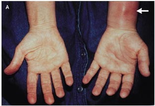
   หญิงอายุ 60 ปี มาตรวจเนื่องจากเจ็บมือซ้ายมา 1 เดือน เมื่อ 3 เดือนก่อน ผู้ป่วยถูกเสี้ยนไม้ตำที่นิ้วหัวแม่มือซ้ายและได้ดึงเสี้ยนออกเองออก ผู้ป่วยได้รับการรักษาด้วยยาปฏิชีวนะและ NSAID แต่อาการกลับเป็นมากขึ้น รอยโรคจากเดิมที่มีการอักเสบและกดเจ็บเฉพาะที่นิ้วหัวแม่มือ กลายเป็นลุกลามเป็นบวมทั้งมือ ข้อมือและแขนซ้ายร่วมกับกดเจ็บและขยับไม่ได้
On examination, the patient appeared healthy and had a temperature of 37.1°C. Her left hand was diffusely swollen, and her fingers were in a semiflexed position. The center of her palm was warm and puffy, and her volar forearm proximal to her left wrist had a red, raised, inflamed area that measured 7 by 5 cm (รูป A, arrow). There was no palpable epitrochlear or axillary adenopathy.
Her white-cell count was 8700/cumm.

1. การวินิจฉัยที่น่าจะเป็นคือ..................................................
2. จะ manage อย่างไรต่อ


Posted by : chpantip , E-mail : (chpantip@medicine.psu.ac.th) ,
Date : 2009-07-29 , Time : 15:40:57 , From IP : 172.29.3.68


ความคิดเห็นที่ : 1


   ขอตอบค่ะ
ในรูป her volar forearm proximal to her left wrist had a red, raised, inflamed area that measured 7 by 5 cm (ตรงลูกศรชี้) คิดว่าเป็น tenosynovitis ค่ะ
เชื้อที่ทำให้เกิด infectious tenosynovitis คือ
o Gonococcus
o Staphylococcus aureus and Streptococcus species are the most common etiologic agents, but infection frequently is mixed (aerobic and anaerobic).
o Pasteurella multocida is common with cat bites; Eikenella corrodens occurs with human bites. However, human and animal bites may have a mixture of aerobic and anaerobic flora.
o Predisposing factors include diabetes mellitus, intravenous (IV) drug abuse, debility, and arteriosclerosis obliterans.
o Mycobacterium species also can cause tenosynovitis, particularly in immunocompromised patients.

ผู้ป่วยเคยถูกเสี้ยนไม้ตำที่นิ้วหัวแม่มือซ้ายเมื่อ 3 เดือนก่อน น่าจะเป็น chronic infectiion ที่เกิดจาก M. tuberculosis หรือ non-tuberculous mycobacterium
ต้องหาเชื้ออื่นๆ เช่น B. pseudomallei, Nocardia spp. และ เชื้อราด้วยค่ะ


Posted by : lara , Date : 2009-08-01 , Time : 14:56:38 , From IP : 172.29.3.68

ความคิดเห็นที่ : 2


   คุณหมอ lara ตอบถูกต้องแล้วค่ะ เก่งมากค่ะ

แพทย์ได้ส่งตรวจ MRI (รูป B)
T1-weighted magnetic resonance image with fat suppression, obtained after the administration of gadolinium, showed enhancement of areas of soft-tissue thickening and swelling around the synovial tendon sheaths, predominantly in the carpal tunnel (central white area); adjacent collections of fluid (long arrows); and cross sections of the flexor tendons (short arrows) and metacarpals (arrowheads); the inset shows a normal wrist.


Posted by : chpantip , Date : 2009-08-04 , Time : 11:51:20 , From IP : 172.29.3.68

ความคิดเห็นที่ : 3


   ได้เจาะดูดเอา fluid ที่ขังอยู่แถว flexor tendons ที่อยู่สูงกว่าข้อมือ ได้หนอง 0.5 มล.
รูป C เป็น สเมียร์ของหนองย้อมด้วย Kinyoun staining
การวินิจฉัยคือ..............................................................


Posted by : chpantip , Date : 2009-08-04 , Time : 11:52:42 , From IP : 172.29.3.68

ความคิดเห็นที่ : 4


   ขอตอบครับ
การวินิจฉัยคือ............
สำหรับ Kinyoun stain เป็นการย้อมเชื้อที่ acid-fast โดยเฉพาะ mycobacterium ซึ่งวิธีจะคล้ายกับ Ziehl-Neelsen stain แต่ทำ slide ให้ร้อน จึงอาจเรียกได้ว่าเป็น “cold” acid-fast stain [Diagnostic Bacteriology: A Study Guide," Margaret A. Bartelt, 2000]
การวินิจฉันคือ mycobacterial infection ซึ่งทำให้เกิดเป็น tenosynovitis ดังกล่าว


Posted by : weeratian , E-mail : (weeratian@hotmail.com) ,
Date : 2009-08-10 , Time : 19:03:25 , From IP : 172.29.11.74


ความคิดเห็นที่ : 5


   *ขอแก้คำผิดครับ
แต่ทำ slide "ไม่" ให้ร้อน


Posted by : weeratian , Date : 2009-08-10 , Time : 19:04:40 , From IP : 172.29.11.74

สงวนสิทธิ์การแสดงความคิดเห็นสำหรับ สมาชิกเท่านั้น