ขอตอบค่ะ
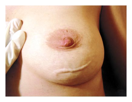
การวินิจฉัย: Mondor disease or sclerosing periphlebitis of the chest wall
Mondor disease is named after Henri Mondor (1885-1960), a French surgeon, who in 1939 was the first to fully describe this condition - namely, a thrombophlebitis generally affecting the thoracoepigastric, lateral thoracic or the superficial epigastric veins. It is an uncommon condition. Incidence rates of 0.84 to 0.96% have been reported. It occurs three times as frequently in women as in men, generally in the age group of 30 to 60 years. The sudden appearance of a rope-like band along the anterior chest wall is characteristic. Erythematous and tender at first, it thereafter changes into a painless tough cord-like strand. Both sides of the chest have the same incidence of involvement. Rarely, the condition is bilateral. Many reasons have been ascribed to its onset and development - strenuous exercise, trauma, infection near the affected vessels, stereotactic vacuum assisted biopsy of the breast, underlying overt or occult malignant disease of the breast, tight clothing, pregnancy and a large pendulous breast.
It has been postulated that direct trauma to the veins, or pressure on the lateral thoracic veins leading to stasis of blood may be the cause. In other cases, the most reasonable explanation is on the basis of repeated movement of the breast along with contracting and relaxing pectoral muscles, which cause stretching and relaxing of the veins. A majority of biopsied lesions show a subcutaneous vein with organizing thrombi and fibrous thickened walls.
Mammographic findings are of a tubular density, along with a sonographic finding of a superficial vessel with or without an intraluminal thrombus. A tubular anechoic structure with multiple areas of narrowing giving a beaded appearance to the vessel and without flow on Doppler Imaging are the typical findings of Mondor disease.
The course of the disease is uneventful. There is no increased risk of blood clots in other veins. The condition subsides within 2-8 weeks, leaving no traces or complications in its aftermath. Treatment is symptomatic and as the discomfort is minor, warm compresses and analgesics will generally suffice.
Thus, while generally being a benign and self-limiting condition, the physician should be aware that Mondor disease might occasionally be the presenting symptom of breast cancer. Appropriate investigations to rule out this possibility must be conducted.
Muthachen M. Linear cord-like lesion on the chest wall
Indian Journal Dermatology, Venereology and Leprology 2006;72: 252.
Posted by : lara , Date : 2009-07-25 , Time : 14:29:34 , From IP : 172.29.3.68
|