ความคิดเห็นทั้งหมด : 5

a 51-YOW with rheumatoid arthritis had swelling and loss of function of right shoulder.


   หญิงอายุ 51 ปีซึ่งเป็น rheumatoid arthritis มานาน 15 ปี มารพ.เนื่องจากไหล่ขวาบวมและใช้งานไม่ได้

1. รูป A คือ Magnetic resonance imaging ของไหล่ของผู้ป่วย พบความผิดปกติอะไรบ้าง
2. จะ manage อย่างไรต่อไป


Posted by : chpantip , E-mail : (chpantip@medicine.psu.ac.th) ,
Date : 2009-07-13 , Time : 11:26:00 , From IP : 172.29.3.68


ความคิดเห็นที่ : 1


   1. รูป A คือ Magnetic resonance imaging ของไหล่ของผู้ป่วย พบความผิดปกติอะไรบ้าง
- จากประวัติผู้ป่วยรายนี้เป็น rheumatoid arthritis มานานซึ่งสัมพันธ์กับการเกิด destruction of head of Rt.humerus
2. จะ manage อย่างไรต่อไป
- ทำ puncture เพื่อดู synovial fluid analysis เพื่อ R/O crystalline disease , infection ออกไป
- CBC,ESR
- เนื่องจากสงสัยว่ามี progressive joint damage เกิดขึ้นแล้ว ควรให้ DMARDs ร่วมกับ glucocorticoid และอาจต้องพิจารณา function ของแขนอีกครั้ง หาก severe functional อาจจำเป็นต้องผ่าตัดร่วมด้วย


Posted by : harder , Date : 2009-07-13 , Time : 18:38:52 , From IP : 172.29.22.43

ความคิดเห็นที่ : 2


   คุณหมอ harder ตอบถูกต้องแล้วค่ะ

MRI of the shoulder revealed severe destruction of the humeral head with an elevated signal of the bone marrow (รูป A).

เจาะข้อไหล่ ได้น้ำสีขาวขุ่น (รูป B) 60 มล.


Posted by : chpantip , Date : 2009-07-15 , Time : 10:47:01 , From IP : 172.29.3.68

ความคิดเห็นที่ : 3


   รูป C คือการตรวจน้ำจากข้อด้วย polarizing microscopy

การวินิจฉัย คือ................................................


Posted by : chpantip , Date : 2009-07-15 , Time : 10:47:34 , From IP : 172.29.3.68

ความคิดเห็นที่ : 4


   จากลักษณะของ synoveal fluid ขาวขุ่นเหมือนน้ำนม(chylous fluid) ไม่พบ wbc มีแต่ crystal เป็นแผ่นสี่เหลี่ยม มีหักที่มุม ที่ค้นได้ น่าจะเป็น cholesterol crystal (5–40 μm. +vely birefringent notched plates) ค่ะ

Posted by : lara , Date : 2009-07-18 , Time : 13:57:01 , From IP : 172.29.3.68

ความคิดเห็นที่ : 5


   คุณหมอ lara ตอบถูกต้องแล้วค่ะ เก่งมากค่ะ

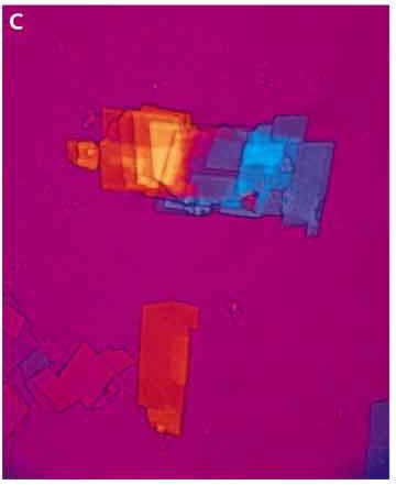
Microscopy of the synovial fluid drawn from this shoulder showed geometric plates with notched corners, pathognomonic of monohydrate cholesterol crystals (รูป C)
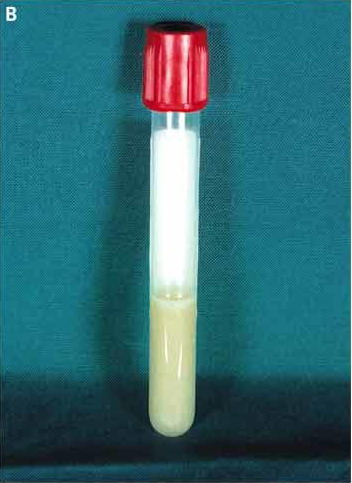
Analysis of the synovial fluid revealed a
-leukocyte count 2100/cumm
-total cholesterol level 7.8 g/L,
-low-density lipoprotein cholesterol level 6.1 g/L
-high-density lipoprotein cholesterol level 1.7 g/L
-triglyceride level 0.83 g/L
plasma lipid profile was normal.

Cholesterol crystals พบได้เป็นครั้งคราวใน synovial fluid ของผู้ป่วย rheumatoid arthritis Crystals เหล่านี้อาจปรากฏใน 2 รูปแบบคือ
1. large, flat, rectangular plates that are negatively birefringent (doubly refracting) with notched corners, ranging from 8 to 100 µm long and consisting of monohydrate cholesterol, or
2. rod-shaped, helical birefringent crystals, ranging from 2 to 20 µm long and consisting of anhydrate cholesterol.

เนื่องจาก large cholesterol plates ถูกกำจัดได้ยาก จึงเข้าใจกันว่า cholesterol crystal ทำให้เกิดข้ออักเสบอย่างต่อเนื่อง.


Reference: Jansen TL, Spoorenberg A. A medical mystery — arthritis.
NEJM 2006; 354 (22): 2375.


Posted by : chpantip , Date : 2009-07-18 , Time : 16:17:38 , From IP : 172.29.3.68

สงวนสิทธิ์การแสดงความคิดเห็นสำหรับ สมาชิกเท่านั้น