ความคิดเห็นทั้งหมด : 3

A 53-YOM had abnormal of his eyes and pain &numbness of left arm


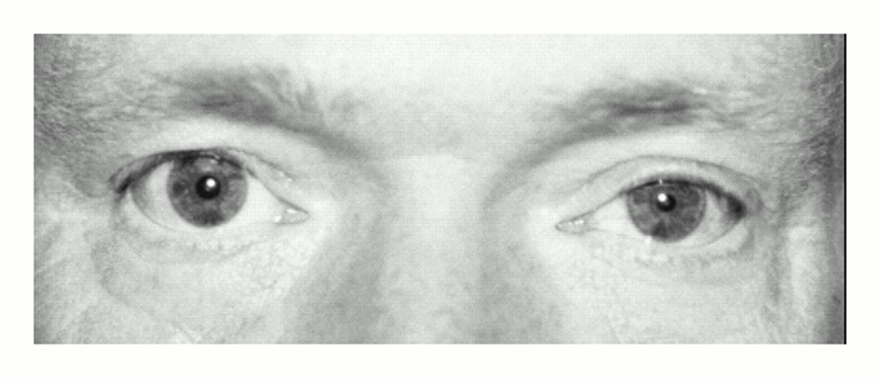
   ชายอายุ 53 ปีถูกส่งตัวมาปรึกษาเนื่องจากมีความผิดปกติของตาซ้ายดังในรูป A
เขาปวดและชาที่แขนซ้ายร่วมกับเหงื่อออกน้อยลงที่หน้าด้านซ้ายมานาน 9 เดือน

1. ความผิดปกติของตาคือ..................................................
2. กลุ่มอาการนี้คือ..............................................................
3. จะ manage อย่างไรต่อไป


Posted by : chpantip , E-mail : (chpantip@medicine.psu.ac.th) ,
Date : 2009-07-03 , Time : 09:52:15 , From IP : 172.29.3.68


ความคิดเห็นที่ : 1


   1. ความผิดปกติของตาคือ miosis + partial ptosis
2. กลุ่มอาการนี้คือ Horner syndrome
3. จะ manage อย่างไรต่อไป >> หา cause ที่ทำให้ damage ของ sympathetic nervous system
- Hx เพิ่มเติมเช่น Hx.Trauma + surgery ก่อนหน้านี้ , ประวัติมะเร็งในครอบครัว เบื่ออาหาร น้ำหนักลด ประวัติแขนขาอ่อนแรงก่อนหน้านี้ ฯลฯ
- confirm Horner syndrome ด้วย Topical cocaine test
- investigate เพิ่มเติมเช่น CXR , CT-scan >> หากประวัติตรวจร่างกายเข้าได้กับ stroke หรือทำ MRI หากสงสัยพวกเนื้องอกต่างๆ


Posted by : harder , Date : 2009-07-03 , Time : 14:11:33 , From IP : 172.29.14.73

ความคิดเห็นที่ : 2


   คุณหมอ harder ตอบถูกแล้วค่ะ

เขามี anisocoria และ left-sided ptosis 1 มม. มานาน 1 เดือน (รูป A). Anisocoria เห็นได้ชัดที่สุดเมื่ออยู่ในที่มืด การตรวจด้วยการหยอดตาทั้ง 2 ข้างด้วย 10% cocaine solution ทำให้เกิดการขยายตัวของ pupil ของตาขวาเท่านั้น

The cocaine test ทำให้วินิจฉัย Horner”s syndrome ได้ เมื่อพบว่า pupil ข้างที่มี sympathetic interruption ไม่ขยายตัว. เนื่องจาก cocaine ปิดกั้น reuptake ของ tonically released norepinephrine จึงทำให้ pupil ข้างที่ไม่มี sympathetic มาเลี้ยง ขยายตัวน้อยหรือไม่ขยายตัวเลย

รูป B คือ T1-weighted gadolinium-enhanced magnetic resonance image of the patient”s chest พบความผิดปกติอะไรบ้าง


Posted by : chpantip , Date : 2009-07-04 , Time : 11:01:48 , From IP : 172.29.3.68

ความคิดเห็นที่ : 3


   T1-weighted gadolinium-enhanced magnetic resonance image of the patient”s chest shows a soft-tissue mass of 4 by 5 by 2.5 cm (solid arrow) at the apex of the left lung adjacent to the T1 vertebral body. This appears in contrast to the normal air-filled apex of the right lung (open arrow).

Biopsy of the mass under computed tomographic guidance revealed a poorly differentiated adenocarcinoma.

ผู้ป่วยได้รับการผ่าตัด left upper lobectomy with chest-wall resection of the first, second, and third ribs.

Horner”s syndrome และอาการชาที่แขนยังคงมีอยู่หลังการผ่าตัด

Reference: Balcer LJ, Galetta SV. Pancoast”s Syndrome. NEJM 1997;337 (19): 1359


Posted by : chpantip , Date : 2009-07-04 , Time : 11:05:07 , From IP : 172.29.3.68

สงวนสิทธิ์การแสดงความคิดเห็นสำหรับ สมาชิกเท่านั้น