ความคิดเห็นทั้งหมด : 5

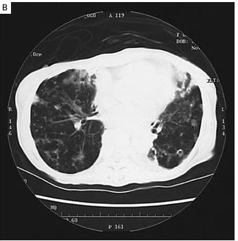
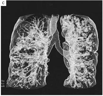
An 81-YOW had abnormal weight loss, hemoptysis and abnorma; CXR


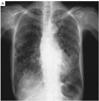
   หญิงอายุ 81 ปีเข้ารับการรักษาในรพ.เนื่องจากน้ำหนักลด (18 กก.ใน 27 เดือน), ไอเป็นเลือด และมีภาพรังสีทรวงอกผิดปกติ

Physical examination demonstrated persistent, coarse rales and diffuse rhonchi throughout the lungs, clubbing, and cyanosis.

1. รูป A คือภาพรังสีทรวงอกของผู้ป่วย พบความผิดปกติอะไรบ้าง
2. การวินิจฉัยคือ..............................................................


Posted by : chpantip , E-mail : (chpantip@medicine.psu.ac.th) ,
Date : 2009-06-30 , Time : 15:22:40 , From IP : 172.29.3.68


ความคิดเห็นที่ : 1


   diffuse reticulopatchy infiltraion with hyperaeration
R/O Bronchioloalveolar carcinoma ครับ


Posted by : patsk120 , E-mail : (patsk120@hotmail.com) ,
Date : 2009-07-01 , Time : 08:59:03 , From IP : 118.173.6.139.adsl.d


ความคิดเห็นที่ : 2


   ขอบคุณคุณหมอ patsk มากค่ะ

Her chest radiograph shows tubular and diffuse granular shadows.(รูปA).

รูป B คือ high-resolution computed tomographic (CT) scan of the lungs (Panel B)


Posted by : chpantip , Date : 2009-07-01 , Time : 10:46:13 , From IP : 172.29.3.68

ความคิดเห็นที่ : 3


   รูป C คือ three-dimensional image of the lungs that was reconstructed from the CT scan

1. ทั้ง 2 รูป พบความผิดปกติอะไรบ้าง
2. การวินิจฉัยคือ.................................


Posted by : chpantip , Date : 2009-07-01 , Time : 10:46:55 , From IP : 172.29.3.68

ความคิดเห็นที่ : 4


   1. ทั้ง 2 รูป พบความผิดปกติอะไรบ้าง
- ลักษณะเป็นแบบ bronchiectasis ที่มี mucoid impaction ที่ปอดทั้ง 2 ข้าง + multiple nodules บางอันเหมือนเป็นโพรงอยู่ข้างในด้วย(cavity)
- รูป C >> เห็นเป็นลักษณะของ saccular ที่ปอดทั้ง 2 ข้าง
2. การวินิจฉัยคือ...
- จากภาพ CXR และประวัติไอเป็นเลือด คิดถึง bronchiectasis ที่อาจมี infection ร่วมด้วย เนื่องจากปอดไม่พบ mass และลักษณะของ CXR ดูเปรอะเหมือนมี infection ร่วมด้วยครับ

*ขออภัย ถ้าใช้ภาษาผิดครับ ความรู้ extern มีน้อย เร่งศึกษา เมื่อจบไปเจ้าจะได้มีวิชา เป็นเครื่องหาช่วยเหลือได้ถูกวิธี * ^ ^


Posted by : harder , Date : 2009-07-03 , Time : 17:01:44 , From IP : 172.29.14.73

ความคิดเห็นที่ : 5


   คุณหมอ harder ตอบได้ดีมากค่ะ

รูป B คือ high-resolution computed tomographic (CT) scan of the lungs demonstrated the presence of bronchiectasis, with ectatic markings; mucoid impaction; multiple small, well-circumscribed pulmonary nodules, some of which had cavities; and granular shadows throughout the lungs.
A three-dimensional image of the lungs that was reconstructed from the CT scan showed saccular and varicose ectatic bronchi (รูป C).

ผู้ป่วยได้รับการตรวจ bronchoscopy ทำ bronchial washings และ sputum cultures ขึ้น Mycobacterium avium complex.

Bronchiectasis พบได้บ่อยในผู้ป่วยที่มีการติดเชื้อ M. avium complex


Reference: Ebihara T, Sasaki H. Bronchiectasis with Mycobacterium avium Complex Infection. NEJM 2002; 346 (18): 1372.


Posted by : chpantip , Date : 2009-07-04 , Time : 10:19:39 , From IP : 172.29.3.68

สงวนสิทธิ์การแสดงความคิดเห็นสำหรับ สมาชิกเท่านั้น