ความคิดเห็นทั้งหมด : 5

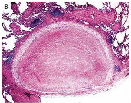
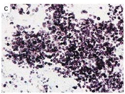
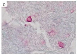
A 48-YOM who underwent pancreas–kidney transplantation had lung nodules


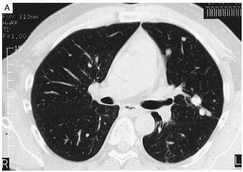
   ผู้ป่วยเบาหวานเพศชายอายุ 48 ปี ได้รับการผ่าตัด pancreas–kidney transplantation สำหรับ end-stage renal disease.
1 เดือนต่อมา เขาได้รับการรักษาสำหรับ rejection และได้รับการตรวจ CT chest ไม่พบ pulmonary infiltrates.
9 เดือนหลัง transplantation เขาเกิดเป็น cryptococcal meningitis. เขาได้รับการรักษาด้วย intravenous amphotericin B–lipid complex, และได้รับ oral fluconazole ต่อสำหรับ long- therapy. serum cryptococcal antigen titer ตอนแรก 1:512 แล้วลดถึงระดับวัดไม่ได้ใน 5 ปี

6 ปีหลัง transplantation เขามี progressive renal-allograft failure ตรวจพบความผิดปกติของภาพรังสีทรวงอก ผู้ป่วยไม่มีไข้หรือ อาการทางปอดใดๆ

CT chest (รูป A) demonstrated 30 variably calcified nodules in the left lung, measuring 1 to 15 ml in diameter.


Posted by : chpantip , E-mail : (chpantip@medicine.psu.ac.th) ,
Date : 2009-06-24 , Time : 10:09:55 , From IP : 172.29.3.68


ความคิดเห็นที่ : 1


   รูป B เป็น thoracoscopic lung biopsy (hematoxylin and eosin, x13) พบความผิดปกติอะไร

Posted by : chpantip , Date : 2009-06-24 , Time : 10:10:54 , From IP : 172.29.3.68

ความคิดเห็นที่ : 2


   รูป C เป็น thoracoscopic lung biopsy (methenamine silver stain, x132) พบความผิดปกติอะไร

Posted by : chpantip , Date : 2009-06-24 , Time : 10:11:26 , From IP : 172.29.3.68

ความคิดเห็นที่ : 3


   รูป D เป็น thoracoscopic lung biopsy (mucicarmine stain, x132) พบความผิดปกติอะไร

การวินิจฉัยคือ.....................................................................


Posted by : chpantip , Date : 2009-06-24 , Time : 10:12:08 , From IP : 172.29.3.68

ความคิดเห็นที่ : 4


   ขอตอบค่ะ
จาก methenamine silver stain พบ yeast จำนวนมาก และ yeast นี้ย้อมติดสี mucicarmine stain
การวินิจฉัย: pulmonary cryptococcosis


Posted by : lara , Date : 2009-06-26 , Time : 14:44:16 , From IP : 172.29.3.68

ความคิดเห็นที่ : 5


   คุณหมอ Lara ตอบถูกแล้วค่ะ

CT chest demonstrated 30 variably calcified nodules in the left lung, measuring 1 to 15 ml in diameter.

Thoracoscopic lung biopsy showed necrotizing granulomas (รูป B; hematoxylin and eosin, x13) containing yeast forms (รูป C; methenamine silver stain, x132). There were both intact and degenerate spores with thick mucin-positive capsules, consistent with the presence of Cryptococcus neoformans (Panel D; mucicarmine stain, x132).

ผู้ป่วยได้รับ kidney transplant ครั้งที่ 2 จาก living related donor.

9 เดือนหลัง transplantation เขายังคงได้รับการรักษาด้วย fluconazole 200 mg/วัน serum cryptococcal antigen titers ยังคงอยู่ในระดับที่วัดไม่ได้

Reference:
Fairhurst RM, Pegues DA. Pulmonary Cryptococcal Granulomas
347 (7): 497.


Posted by : chpantip , Date : 2009-06-27 , Time : 15:30:31 , From IP : 172.29.3.68

สงวนสิทธิ์การแสดงความคิดเห็นสำหรับ สมาชิกเท่านั้น