ความคิดเห็นทั้งหมด : 2

A 34-year-old woman had back pain


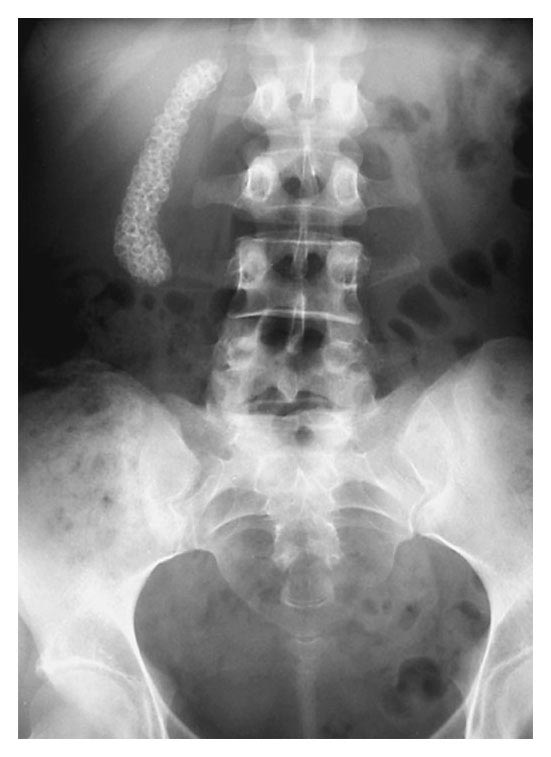
   หญิงอายุ 34 ปีซึ่งสุขภาพดี มารพ.เนื่องจากปวดหลัง
ผลการตรวจร่างกายและ lab อยู่ในเกณฑ์ปกติ.
แพทย์ได้ส่งผู้ป่วยไปถ่ายภาพรังสีของ lumbar spine.

1. พบความผิดปกติอะไรบ้างในภาพรังสีของ lumbar spine.
2. การวินิจฉัยคือ...............................................


Posted by : chpantip , E-mail : (chpantip@medicine.psu.ac.th) ,
Date : 2009-05-08 , Time : 15:00:10 , From IP : 172.29.3.68


ความคิดเห็นที่ : 1


   ขอตอบค่ะ

ในรูปมี stones จำนวนมากอยู่ใน structure ที่ RUQ ซึ่งน่าจะเป็น gallbladder ที่ยาวลงมา

การวินิจฉัยคือ gallbladder stones ค่ะ


Posted by : lara , Date : 2009-05-12 , Time : 15:54:04 , From IP : 172.29.3.68

ความคิดเห็นที่ : 2


   คุณหมอ Lara ตอบถูกต้องแล้วค่ะ

The radiograph showed multiple faceted stones outlining the contours of the gallbladder.

Ultrasonography of the upper abdomen (ไม่มีรูป) showed multiple gallstones with acoustic shadowing throughout the gallbladder, which did not contain visible bile.

ผู้ป่วยได้รับการทำ laparoscopic surgery เพื่อเอานิ่วออกจากถุงน้ำดี มีนิ่วจำนวนมากอยู่ภายในถุงน้ำดี ผู้ป่วยหายเป็นปกติ

Reference:
Yekeler E and Akyol Y. Cholelithiasis. N Engl J Med 2004;351 (22): 2318.


Posted by : chpantip , Date : 2009-05-15 , Time : 15:33:45 , From IP : 172.29.3.68

สงวนสิทธิ์การแสดงความคิดเห็นสำหรับ สมาชิกเท่านั้น