ความคิดเห็นทั้งหมด : 5

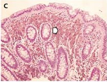
A 60-YOW with a family history of CA colon underwent surveillance colonoscopy


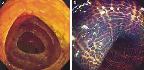
   หญิงอายุ 60 ปีได้มารับการตรวจ surveillance colonoscopy. มีประวัติคนในครอบครัวของเธอป่วยเป็น colon cancer และเป็น adenomatous colorectal polyps.
เธอได้รับการตรวจ endoscopy หลังสุดเมื่อ 3 ปีก่อนซึ่งพบว่ามี polyp 2 อันและ mucosa ของ colon ปกติ (รูป A).

การตรวจครั้งนี้พบลักษณะของ mucosa ของ colon ทั้งหมดดังในรูป B ไม่พบความผิดปกติอื่นๆ
ส่วนรูป C เป็น biopsy ของ colon

1. การวินิจฉัยคือ................................................
2. จะถามประวัติอะไรจากผู้ป่วย
3. จะ manage อย่างไร


Posted by : chpantip , E-mail : (chpantip@medicine.psu.ac.th) ,
Date : 2009-04-06 , Time : 10:49:42 , From IP : 172.29.3.68


ความคิดเห็นที่ : 1


   รูป C เป็น biopsy ของ colon

Posted by : chpantip , Date : 2009-04-06 , Time : 10:50:39 , From IP : 172.29.3.68

ความคิดเห็นที่ : 2


   ขอตอบว่า
1. การวินิจฉัยคือ melanosis coli ซึ่งมี pigment สีน้ำตาลดำสะสมอยู่ใน macrophage ในชั้น lamina propria ของลำไส้ใหญ่

2. ถามผู้ป่วยเรื่องการกินยาถ่ายประเภท senna คือ senokot ค่ะ

3. ให้หยุด senokot


Posted by : lara , Date : 2009-04-08 , Time : 14:21:33 , From IP : 172.29.3.68

ความคิดเห็นที่ : 3


   คุณหมอ lara ตอบถูกต้องแล้วค่ะ

ผู้ป่วยมีท้องผูกเป็นมานานเป็นปี และเป็นมากขึ้น ต้องกิน senna (ซึ่งเป็นยาระบายที่มี anthraquinone) ทุกวันจึงจะถ่าย

Colonoscopy revealed diffuse dark brown and black pigmentation throughout her colon, which is consistent with the presence of melanosis coli (รูป B).
Biopsies of the affected areas: pigment in macrophages of the lamina propria, without any evidence of inflammation or cancer (รูป C).


Posted by : chpantip , Date : 2009-04-09 , Time : 08:14:36 , From IP : 172.29.3.68

ความคิดเห็นที่ : 4


   Melanosis coli เป็น dark brown pigmentation ของ colon ซึ่งเกิดขึ้นจากการกินยาถ่ายที่มี anthraquinone เช่น senna. Melanosis coli เกิดขึ้นได้ภายในเวลา 2-3 เดือนของการใช้ยานี้ และหายไปได้หลังจากหยุดยา 2-3 เดือน.

Anthraquinones มี direct toxic effect ต่อ epithelial cells ของ colon โดยมีผลทำให้เกิดการสร้าง lipofuscin ซึ่งเป็น pigment สีน้ำตาลดำ อยู่ใน macrophages ใน melanosis coli (รูป C, ลูกศร). cecum และ rectum เป็นตำแหน่งที่พบ melanosis coli บ่อยที่สุด แต่ก็สามารถพบได้ทั่วลำไส้ใหญ่.

Melanosis coli เป็น benign lesion เพราะไม่พบว่ามีความสัมพันธ์กับมะเร็งของลำไส้ใหญ่

Ref. Ahmed S, et al. Melanosis Coli. NEJM 2003; 349: 1349.


Posted by : chpantip , Date : 2009-04-09 , Time : 08:15:58 , From IP : 172.29.3.68

ความคิดเห็นที่ : 5


   มี comment เกี่ยวกับ pathophysiological mechanism ของ melanosis coli และ terminology

The pigmentation is triggered by deposits of apoptotic cells, which are ingested by adjacent macrophages within the epithelial tissue.2 Those macrophages migrate in the lamina propria, where lysosomes convert the remains of the cells into lipofuscin pigment. Thus, the term "melanosis" may be misleading, since the pigment is not melanin. As a reference to the dark color of the pigment, the term "nigrosis" coli (from the Latin word "niger," meaning black) would be more appropriate.2 In addition to its association with anthraquinones, nigrosis coli may appear during long-term use (use for a period of more than four months) of bisacodyl. It spontaneously declines within a few years after discontinuation of the laxative.

Mennecier D, Vergeau B. N Engl J Med 2004; 350:197.

เพิ่มเติม term "melanosis coli" was coined in 1857 by the German pathologist Dr. Rudolf Virchow, who is considered to be the father of modern pathology.


Posted by : chpantip , Date : 2009-04-10 , Time : 11:33:28 , From IP : 172.29.3.68

สงวนสิทธิ์การแสดงความคิดเห็นสำหรับ สมาชิกเท่านั้น