ความคิดเห็นทั้งหมด : 5

A 71-YOW with bronchiectasis had dyspnea and cough for 2 wks


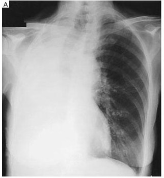
   หญิงอายุ 71 ปีซึ่งมี bronchiectasis อยู่เดิมได้เข้ารับการรักษาในรพ.เนื่องจากหอบเหนื่อยและไอมีเสมหะ เสมหะสีเหลือง เหนียวและมีปริมาณน้อยมานาน 2 สัปดาห์. ผู้ป่วยได้รับยาปฏิชีวนะจากแพทย์ที่คลินิกมาก่อนแต่อาการไม่ดีขึ้น.
ผู้ป่วยหอบเหนื่อยมากขึ้น 1 วันก่อนเข้ารับการรักษาในรพ.

ในรูป A เป็น chest radiography ถ่ายเมื่อแรกรับเข้ารพ. พบความผิดปกติอะไรบ้าง (CXR ของผู้ป่วยเมื่อ 2 สัปดาห์ก่อนอยู่ในเกณฑ์ปกติ)


Posted by : chpantip , E-mail : (chpantip@medicine.psu.ac.th) ,
Date : 2009-04-03 , Time : 08:27:22 , From IP : 172.29.3.68


ความคิดเห็นที่ : 1


   Large Rt.lung patch, Trachea shift to the right.

Posted by : harder , Date : 2009-04-04 , Time : 16:38:27 , From IP : 114.128.146-153.dyna

ความคิดเห็นที่ : 2


   findings
- Trachea and mediastinum shift to the right
- Soft tissue density entire right hemithorax
- decrease ICS of right hemithorax
IMP: Right lung atelectasis most likely from secretion obstruction


Posted by : hata_o , Date : 2009-04-05 , Time : 10:04:36 , From IP : 172.29.22.44

ความคิดเห็นที่ : 3


   ขอลองตอบครับ
ที่เห็นเป็น homogenous opacity ที่ปอดด้านขวา ร่วมกับ มี signs ของ volume loss ของปอดด้านขวา คือ มี trachea และ mediastinum shift ไปด้านขวา และ intercostals space ด้านขวาลดลง ดังนั้นจึงคิดว่าน่าจะเป็น “complete right lung atelectasis” ส่วนสาเหตุนั้นคิดว่าหากผู้ป่วยมี underlying disease เดิมคือ brochiectasis สาเหตุน่าจะมาจากการอุดกลั้น airways ด้วย sputum ที่ไม่สามารถขับออกมาได้ตามปกติเหมือนคนทั่วไป หรือ อาจเกิดจาก clotted blood ซึ่งผู้ป่วย bronchiectasis มี hemoptysis ได้


Posted by : weeratian , Date : 2009-04-05 , Time : 14:43:17 , From IP : 125.24.95.171.adsl.d

ความคิดเห็นที่ : 4


   คุณหมอ harder,คุณหมอ hata-0 และ คุณหมอ Weeratian ตอบถูกต้องแล้วค่ะ

Chest radiography showed opacification of and volume loss in the right lung (Panel A). A chest radiograph obtained two weeks earlier had been normal except for changes indicative of bronchiectasis.

Chest physiotherapy ไม่ได้ช่วยให้มีเสมหะออกมากขึ้น

Fiberoptic bronchoscopy: a large mucous plug completely occluding the right main bronchus (รูป B).


Posted by : chpantip , Date : 2009-04-05 , Time : 16:18:03 , From IP : 172.29.3.68

ความคิดเห็นที่ : 5


   ต้องใช้ rigid bronchoscopy เพื่อเอา mucous plug ออกมา. ไม่มี endobronchial lesionใดๆ. ปอดข้างขวาขยายตัวได้เต็มที่หลังเอา plug ออกไป (รูป C). ผู้ป่วยหายใจได้ดีขึ้นมากทันที.

ผลการเพาะเชื้อในเสมหะ: normal respiratory tract flora.

Reference: Nair SR. Mucous plug in the bronchus causing lung collapse
NEJM 2002; 347 (14): 1079.


Posted by : chpantip , Date : 2009-04-05 , Time : 16:19:46 , From IP : 172.29.3.68

สงวนสิทธิ์การแสดงความคิดเห็นสำหรับ สมาชิกเท่านั้น