ความคิดเห็นทั้งหมด : 4
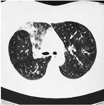
หญิงอายุ 45 ปี มี slowly resolving pneumonia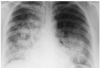 หญิงอายุ 45 ปี มารพ,ด้วยเรื่อง pneumonia. ผู้ป่วยมีประวัติสูบบุหรี่ Chest radiograph: มี consolidation ที่ right upper lung. ผู้ป่วยได้รับยาปฏิชีวนะ อาการต่างๆของผู้ป่วยดีขึ้นจนเเป็นปกติ. อีก 1 เดือนต่อมา แพทย์ได้ส่งตรวจ chest radiography (รูป A) และ high-resolution computed tomographic scan of the thorax (รูป B). 1. พบความผิดปกติอะไรบ้าง 2. การวินิจฉัยที่น่าจะเป็นคือ........................... Posted by : chpantip , E-mail : (chpantip@medicine.psu.ac.th) , Date : 2009-04-01 , Time : 08:24:22 , From IP : 172.29.3.68 |