ความคิดเห็นทั้งหมด : 3

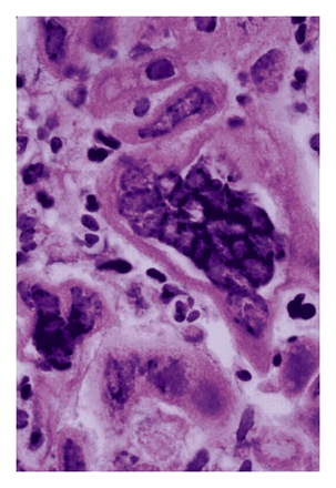
หญิง47 ปี เพลีย คลื่นไส้ อาเจียน ปวดท้องมา 2 วัน และมีอาเจียนเป็นสีดำ


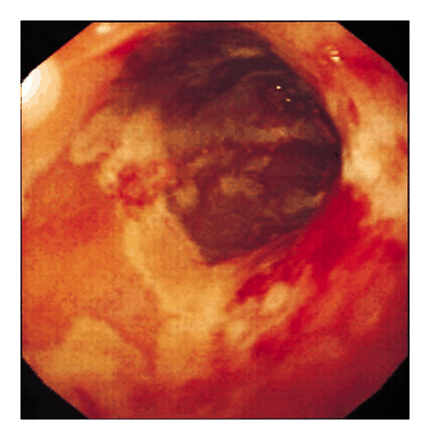
   หญิงอายุ 47 ปีซึ่งมีประวัติความดันโลหิตสูงและดื่มเหล้าจัด มีอาการเพลีย คลื่นไส้ อาเจียนและปวดท้องมา 2 วัน.
แรกรับเข้ารพ. เธออาเจียนเป็น coffee-grounds material.
BP ท่านั่ง 80/50 mm Hg.
Examination revealed no fever or lesions of the mouth. The patient had epigastric tenderness, but the bowel sounds were normal and there was no rebound tenderness.

ผลเลือด: serum amylase level 244 U per liter, serum lipase level 5922, calcium level 3.1 mmol per liter, และ serum bicarbonate level 19 mmol per liter.

ผู้ป่วยได้รับการรักษาด้วย intravenous fluids, ranitidine, pethidine และ bowel rest ทำให้อาการต่างๆดีขึ้น.

เนื่องจากเธออาเจียนเป็นเลือด จึงได้รับการตรวจ esophagogastroduodenoscopy.
EGD scopy revealed severe esophagitis and gastritis. There were inflammatory exudates, ulcerations, and associated granulation tissue in the proximal, middle, and distal portions of the esophagus (รูป A).

1. รูป B คือ esophageal biopsy พบความผิดปกติอะไรบ้าง
2. การวินิจฉัยของ esophagitis คือ...................................


Posted by : chpantip , E-mail : (chpantip@medicine.psu.ac.th) ,
Date : 2009-02-25 , Time : 08:29:41 , From IP : 172.29.3.68


ความคิดเห็นที่ : 1


   รูป B: histology of esophageal biopsy

Posted by : chpantip , Date : 2009-02-25 , Time : 08:31:17 , From IP : 172.29.3.68

ความคิดเห็นที่ : 2


   ขอตอบครับ
-->ข้อที่ 1 รูป B คือ esophageal biopsy พบความผิดปกติอะไรบ้าง?
รูปนี้คล้ายกับ slide ที่เห็นบน ward ตอนสะกิด lesion ของผู้ป่วย HSV บริเวณริมฝีปากครับ
เคยได้รับความรู้จากรุ่นพี่ว่า เป็นลักษณะ "multinucleated giant cell"
ค้นคว้าเพิ่มเติมได้ความว่า:
สำหรับ HSV นั้น histologic examination จะมีลักษณะเป็น ballooning degeneration,
multinucleated giant cells, and prominent intranuclear Cowdry type A inclusion bodies
คิดเป็น 55-70% ของ biopsy specimens.
ทั้งนี้แตกต่างจาก CMV คือ histopathologic examination จะเป็น enlarged endothelial cells or
fibroblasts containing large, dense intranuclear inclusion.
-->2. การวินิจฉัยของ esophagitis คือ................................... ?
สำหรับ esophagitis นั้นมีสาเหตุจาก noninfectious conditions มากที่สุด และ GERD ก็เป็นสาเหตุ
ของ esophagitis มากที่สุด แต่หากเป็น esophageal infection นั้นจะพบในผู้ป่วยที่มี impaired immunity
เช่น ได้รับ cancer chemotherapy, tranplantation หรือ HIV (แต่บางครั้งก็พบใน healthy person)
เชื้อที่เป็นสาเหตุได้แก่ 1)Candidiasis 2)Cytomegalovirus 3)Herpes simplex virus 4)HIV infection, acute
และส่วนหนึ่งของ esophagitis ก็ไม่ทราบสาเหตุ (aphthous (idiopathic) ulceration of the esophagus)
ด้วยอาการแล้ว เราไม่สามารถแยกการติดเชื้อ candida, CMV, HSV ออกจากกันได้ อีกทั้ง gross appearance
ก็แยกกันยาก แม้ว่าอาจจะมีบางลักษณะที่พอบอกได้เช่น CMV lesion เป็น "punched-out" ulcers ในขณะที่
HSV มีลักษณะเป็น "Volcano ulcer"

Histopathologic examination จึงเป็นเครื่องมือวินิจฉัยที่ดีที่สุด(ภาพ 2) จึงขอตอบว่า
--> Herpes Simplex Virus Esophogitis
ref: Mandell, Bennett, & Dolin: Principles and Practice of Infectious Diseases, 6th ed.
(Chapter 90 – Esophagitis By PAUL S. GRAMAN)


Posted by : weeratian , E-mail : (weeratian@hotmail.com) ,
Date : 2009-02-25 , Time : 23:25:39 , From IP : 172.29.22.75


ความคิดเห็นที่ : 3


   คุณหมอ Weeratian ตอบได้ถูกต้องแล้วค่ะ ขอบคุณที่ให้รายละเอียดมาด้วย

Histology of esophageal biopsy demonstrated multinucleated giant cells consistent with the presence of herpetic esophagitis (รูป B; hematoxylin and eosin, x1000).

ผู้ป่วยไม่มีอาการของ esophagitis หรือ dyspepsia.
ตรวจ HIV antibody ได้ผลลบ และไม่พบหลักฐานของ immunodeficiency state ใดๆ.

เธอได้รับการรักษาด้วย oral acyclovir นาน 14 วันและยังอยู่รพ.อีกหลายสัปดาห์เพราะมีภาวะแทรกซ้อนของ pancreatitis.

จากการตรวจตามนัดที่ 6 เดือน เธอไม่มีอาการปวดท้อง กลืนลำบาก หรือหอบเหนื่อย และกินอาหารได้เป็นปกติ.

รูปและเรื่องจาก Urbano FL. NEJM 1999;340 (16): 1254.


Posted by : chpantip , Date : 2009-03-02 , Time : 07:59:58 , From IP : 172.29.3.68

สงวนสิทธิ์การแสดงความคิดเห็นสำหรับ สมาชิกเท่านั้น