ความคิดเห็นทั้งหมด : 4

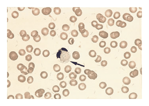
ชาย 41 ปี ปวดท้อง ถ่ายอุจจาระเหลวและมีไข้ 2 วัน


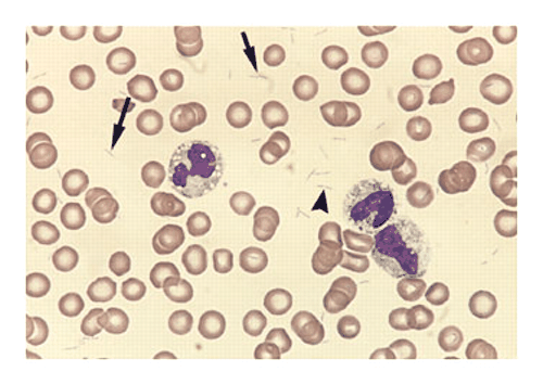
   ชายอายุ 41 ปีมีประวัติปวดท้อง ถ่ายอุจจาระเหลวและมีไข้มานาน 2 วัน

T 40°C, BP 139/59 mm Hg, and pulse rate 100 beats per minute.
Physical examination revealed abdominal distention, diffuse tenderness, decreased bowel sounds, and poor dentition but was otherwise unremarkable.
Lab: HB 9.4 g/dL, white-cell count of 11,700 /cumm and
a platelet count 1,000/cumm.

ในรูปทั้ง 3 รูป เป็น peripheral-blood smear ของผู้ป่วย รูป A และ B เป็นการย้อมสี May–Grünwald–Giemsa ส่วนรูป C เป็น Gram stain

1. ปัญหาของผู้ป่วยรายนี้มีอะไรบ้าง
2. พบความผิดปกติอะไรในรูปทั้ง 3 รูป
3. การวินิจฉัยเบื้องต้นคือ..............................................
4. จะ manage อย่างไร


Posted by : chpantip , E-mail : (chpantip@medicine.psu.ac.th) ,
Date : 2009-02-23 , Time : 08:14:54 , From IP : 172.29.3.68


ความคิดเห็นที่ : 1


   รูป B peripheral-blood smear ของผู้ป่วยย้อมสี May–Grünwald–Giemsa

Posted by : chpantip , Date : 2009-02-23 , Time : 08:16:11 , From IP : 172.29.3.68

ความคิดเห็นที่ : 2


   รูป C peripheral-blood smear ของผู้ป่วยย้อม Gram stain

Posted by : chpantip , Date : 2009-02-23 , Time : 08:17:33 , From IP : 172.29.3.68

ความคิดเห็นที่ : 3


   ขอลองตอบครับ
1. ปัญหาของผู้ป่วยรายนี้มีอะไรบ้าง
1.fever with abdominal pain and diarrhea for 2 days
2.abdomianl distention
3.SIRS with very low platelet
4.poor dentition
2. พบความผิดปกติอะไรในรูปทั้ง 3 รูป
พบ pathogen เป็น Gram-negative rod และ รูป B เห็นใน neutrophil
3. การวินิจฉัยเบื้องต้นคือ Septicemia with abdominal distention(painful)
4. จะ manage อย่างไร
คิดว่าสาเหตุน่าจะมาจาก Gastrointestinal tract มากที่สุดเนื่องจาก
ผู้ป่วยมีอาการทาง GI คือ abdominal pain/distention และ diarrhea
คิดว่าควรส่ง imaging เพื่อดูพยาธิสภาพใน abdomen หากเป็น U/S
ก็จะดีเพราะผู้ป่วยต้องการการวินิจฉัยอย่างรวดเร็ว แต่หากเป็น CT ก็จะได้
รายละเอียดมากกว่า โดยเฉพาะ lesion อยู่บริเวณ retroperitoneal
อย่างที่สองก็ควรเป็น empiric antibiotics


Posted by : weeratian , E-mail : (weeratian@hotmail.com) ,
Date : 2009-02-25 , Time : 23:56:40 , From IP : 172.29.22.75


ความคิดเห็นที่ : 4


   คุณหมอ Weeratian ตอบได้ถูกต้องแล้วค่ะ เก่งมากๆ

จาก blood smear ทั้ง 3 รูป พบ bacilli จำนวนมาก ทั้ง 3 รูป. Bacilli เหล่านี้ยาว 2 - 4 µm (arrows) และส่วนใหญ่อยู่เป็นคู่ (arrowhead) (รูป A; May–Grünwald–Giemsa, x790). อยู่นอกเซลล์เป็นส่วนใหญ่ แต่มีบางตัวที่ถูกกินโดย neutrophils (arrow ในรูป B; May–Grünwald–Giemsa, x790). rods เหล่านี้เป็น gram-negative (arrows ในรูป C; Gram stain, x790).

เมื่อแรกรับได้ทำ blood cultures (BacT/Alert, Organon Teknika)ไป 6 ขวด ผลขึ้น Capnocytophaga canimorsus ทั้ง 6 ขวด.

ผู้ป่วยรายนี้น่าจะมี source ของ bacteremia อยู่ที่ odontogenic infection

Capnocytophaga canimorsus (formerly described as dysgonic fermenter type 2 by the Centers for Disease Control) เป็น long, thin, fusiform, gram-negative rods, often arranged in end-to-end pairs. C. canimorsus can grow under microaerophilic conditions and is indole-negative. ต้องแยกจาก Fusobacterium necrophorum ซึ่งมีลักษณะเหมือนกัน เป็นเชื้อในช่องปากเหมือนกัน แยกจากกันได้โดยการเพาะเชื้อเพราะ F. necrophorum iเป็นindole-positive และ strictly anaerobic.

รูปและเรื่องจาก Alberio L. NEJM 1998 ; 339 (25): 1827


Posted by : chpantip , Date : 2009-03-02 , Time : 08:27:22 , From IP : 172.29.3.68

สงวนสิทธิ์การแสดงความคิดเห็นสำหรับ สมาชิกเท่านั้น