ความคิดเห็นทั้งหมด : 3

ชายอายุ 47 ปี เจ็บหน้าอกมา 1 สัปดาห์
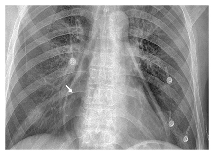


   ชายอายุ 47 ปี มาที่ ER เนื่องจากมีอาการเจ็บหน้าอกมา 1 สัปดาห์
PE: He was hemodynamically stable. The physical examination was unremarkable.

Complete blood count: 27,000 leukocytes per cubic millimeter.

1. Chest radiograph ของผู้ป่วยพบความผิดปกติอะไรบ้าง
2. โปรดให้การวินิจฉัย
3. จะ manage ผู้ป่วยอย่างไรต่อไป


Posted by : chpantip , E-mail : (chpantip@medicine.psu.ac.th) ,
Date : 2008-11-27 , Time : 11:17:16 , From IP : 172.29.3.68


ความคิดเห็นที่ : 1


   1. Chest radiograph ของผู้ป่วยพบความผิดปกติอะไรบ้าง
- There were two obvious radiolucent areas along both heart borders

2. โปรดให้การวินิจฉัย
ผู้ป่วยเป็นชายวัยกลางคน มีอาการเจ็บหน้าอกมา 1 สัปดาห์ ไม่ได้ให้ประวัติ trauma หรือประวัิติที่บ่งบอกถึง pneumothorax หรือ pneumomediastinum ภาพเอกซเรย์เห็น air อยู่ที่ heart border ทั้ง 2 ข้างชัดเจน สรุปว่า Dx : Pneumopericardium

3. จะ manage ผู้ป่วยอย่างไรต่อไป
- ซักประวัติเพิ่มเติม เช่น เรื่อง Trauma , Infection , เคยเป็นมาก่อนหรือไม่ , อาการอื่นๆเช่นไอ สำลัก , ประวัติผ่าตัดหรือรักษามาก่อนหน้านี้ เป็นต้น
- เนื่องจากผู้ป่วยมีไข้ร่วมด้วย ควรส่ง investigate เพิ่มเติม เช่น CT-scan เพื่อหาสาเหตุที่อาจทำให้ติดเชื้อเพิ่มเติม เช่น Fistula กับ esophagus เป็นต้น
- ให้ systematic ATB ร่วมกับใส่ catheter หรือ needle drainage ที่ pericardial space เพื่อระบายอากาศที่ค้างอยู่ออกมา ระวังเรื่อง Cardiac tamponade ด้วยครับ

คิดว่าคงประมาณนี้นะครับ รอความเห็นเพิ่มเติมครับ


Posted by : harder , Date : 2008-11-29 , Time : 17:29:13 , From IP : 172.29.22.43

ความคิดเห็นที่ : 2


   คุณหมอ harder ตอบได้ดีมากค่ะ อาจารย์และ resident ภาคฯอายุรศาสตร์หลายๆ ท่านฝากชมมาด้วยค่ะ

CXR: pneumopericardium (ลูกศร) without evidence of pneumothorax or pneumomediastinum.

Computed tomographic scan of the chest confirmed the diagnosis of pneumopericardium and showed circumferential wall thickening of the distal esophagus with an associated esophagopericardial fistula.

Esophagoscopy revealed a deep esophageal ulcer 4.5 cm in diameter, 36 cm from the incisors; the pericardial cavity and the lining of the columnar epithelium could be directly visualized to the distal 10 cm of the esophagus.

Esophageal-biopsy specimens: intestinal metaplasia with inflammation and high-grade dysplasia.

การวินิจฉัยโรค คือ Barrett esophagus.

ผู้ป่วยได้รับการผ่าตัดโดยทำ lavage และ drainage pericardium ใส่ pericardial patch และ esophageal stent และทำ jejunostomy.
หลังผ่าตัดผู้ป่วยไม่มีภาวะแทรกซ้อนใดๆ การติดตามที่ 1 ปีต่อมา ผู้ป่วยสบายดี


Posted by : chpantip , E-mail : (chpantip@medicine.psu.ac.th) ,
Date : 2008-12-06 , Time : 08:51:08 , From IP : 172.29.3.68


ความคิดเห็นที่ : 3


   เรื่องและรูป จาก
Karoui M, Bucur PO. N Engl J Med 2008;359:e16


Posted by : chpantip , E-mail : (chpantip@medicine.psu.ac.th) ,
Date : 2008-12-06 , Time : 08:53:18 , From IP : 172.29.3.68


สงวนสิทธิ์การแสดงความคิดเห็นสำหรับ สมาชิกเท่านั้น