ความคิดเห็นทั้งหมด : 5

A 33 YOM had white pupil with eosinophilia


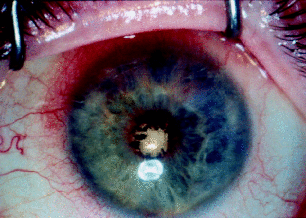
   ชายอายุ 33 ปี มี mental retardation ได้ถูกส่งตัวมาเพื่อ evaluation ตาซ้ายซึ่งแดงและมีจุดขาวที่รูม่านตามานาน 2 เดือน ไม่มีประวัติของภยันตรายต่อตาหรือมีการติดเชื้อใดๆ มาก่อน แม่สังเกตว่า เขามีไข้ต่ำๆ เป็นๆหายๆ

At examination, the patient was unable to fixate and follow with his left eye. His left pupil was not reactive to light and was noted to be leukokoric. The findings of an evaluation of the right eye were normal. The left eye was hypotonous (intraocular pressure, <10 mm Hg) with a hyperemic conjunctiva, shallow anterior chamber, and white pupillary membrane (figure 1).


Posted by : chpantip , E-mail : (chpantip@medicine.psu.ac.th) ,
Date : 2008-11-20 , Time : 11:38:56 , From IP : 172.29.3.68


ความคิดเห็นที่ : 1


   Orbital ultrasound: total retinal detachment with underlying dense subretinal opacities and possible fluid behind both optic nerves, raising suspicions of increased intracranial pressure (figure 2; an orbital ultrasound of his healthy right eye is shown for comparison).
A sample of aqueous fluid was sent for analysis.
ผู้ป่วยได้รับการรักษาในรพ.เพื่อการดูแลรักษาเพราะกลัวจะมี increased intracranial pressure.

เมื่อแรกรับ ผู้ป่วยไม่มีไข้ vital sign ปกติ
PE: a well-developed, well-nourished male subject in no apparent distress. No palpable lymphadenopathy, oral lesions, abdominal tenderness, hepatosplenomegaly, peripheral edema, or rashes were found.
Neurological examination: normal strength and range of motion in all 4 extremities, with no gait disturbances.

Laboratory test results were notable for a marked leukocytosis (21,520 cells/mm3) and eosinophilia (12,912 cells/mm3), as well as an elevated alkaline phosphatase level of 294 IU/L.

Gram staining of the aqueous fluid were negative, and the findings of a cytological examination were unremarkable.

Blood, urine, and stool cultures were sterile.

A CT scan of the brain: no acute intracranial abnormalities.

Analysis of CSF samples showed a normal opening pressure and 4 mononuclear cells/mm3, a glucose level of 53 mg/dL, a protein level of 14 mg/dL, and no eosinophilia;
cultures of CSF samples showed no growth.


Posted by : chpantip , E-mail : (chpantip@medicine.psu.ac.th) ,
Date : 2008-11-20 , Time : 11:50:37 , From IP : 172.29.3.68


ความคิดเห็นที่ : 2


   A CT scan of the thorax revealed multiple pleural-based pulmonary nodules (figure 3).

การวินิจฉัยโรคที่น่าจะเป็นมากที่สุดคืออะไร


Posted by : chpantip , E-mail : (chpantip@medicine.psu.ac.th) ,
Date : 2008-11-20 , Time : 11:52:16 , From IP : 172.29.3.68


ความคิดเห็นที่ : 3


   จากผล lab => Marked leukocytosis (21,520 cells/mm3) and eosinophilia (12,912 cells/mm3)

เหมือนจะมีการติดเชื้อจำพวก Parasitic ครับ และ parasite ที่เกี่ยวข้องกะตา ที่เคยได้ยินมาคือ Ocular larva migran ซึ่งเกิดจากการติดเชื้อปรสิต Toxocara จากอึสุนัขครับ

http://www.vcharkarn.com/vnews/151630

อ้างอิงจากเวปไซต์วิชาการ.คอมนะครับ เวปไซต์นี้ดีมากๆเลยครับ มีความรู้เยอะแยะมาก ลองเข้าไปเยี่ยมชมดูนะครับ แต่ส่วนใหญ่เวปไซต์นี้จะเน้นไปที่ Physics เป็นส่วนใหญ่ครับ เพราะ webmaster จบเกี่ยวกะฟิสิกส์ินิวเคลียร์จาก Oxford university ครับ อิอิ


Posted by : harder , Date : 2008-11-21 , Time : 15:10:29 , From IP : 172.29.22.43

ความคิดเห็นที่ : 4


   ข้อมูลเพิ่มเติมนิดหน่อยครับ เป็นของสัตวแพทย์ แต่แพทย์ก็อ่านเพิ่มเติมได้ครับ

"อัตราความชุกของการติดเชื้อปรสิตในสุนัขจรจัดในเขตกรุงเทพมหานครและปริมณฑล"

http://www.bsru.ac.th/~sci/document/Book2_7.pdf


Posted by : harder , Date : 2008-11-21 , Time : 15:12:25 , From IP : 172.29.22.43

ความคิดเห็นที่ : 5


   คุณหมอ harder ตอบถูกต้องแล้วค่ะ เก่งมากค่ะ

Figure 1. His left eye under an operating microscope showing injected conjunctival vessels with a white pupillary membrane.

Figure 2. Orbital ultrasound of his left eye showing a funnel-shaped total retinal detachment. Arrows indicate edges of retinal detachment. The opacities under the edges of the detachment represent subretinal fluid. The inset shows the healthy right eye, with a clear vitreous cavity and no retinal detachment.
Figure 3. CT scan of a section of his lung showing multiple pleural-based pulmonary nodules.

Diagnosis: Ocular and visceral larva migrans secondary to Toxocara canis infection.

ผล serological test: strongly positive for Toxocara canis, with a titer of 1:256.

จากผลการตรวจตา (รูป 1 และ 2), hypereosinophilia, pulmonary nodules (รูป 3) และ positive serological test results ผู้ป่วยจึงได้รับการวินิจฉัยว่าเป็น ocular and visceral larva migrans due to Toxocara canis การพยากรณ์โรคของตาไม่ดีเนื่องจากมี total retinal detachment เนื่องจากเขามีรอยโรคที่ปอดด้วย จึงได้รับการรักษาด้วย albendazole และprednisolone น่าเสียดายที่ผู้ป่วยไม่ได้มารับการรักษาต่อ

ประวัติเพิ่มเติม เขาไม่ได้เดินทางไปที่ใดมาก่อน ที่บ้านเลี้ยงสุนัข 2 ตัว


Posted by : chpantip , E-mail : (chpantip@medicine.psu.ac.th) ,
Date : 2008-11-25 , Time : 11:06:34 , From IP : 172.29.3.68


สงวนสิทธิ์การแสดงความคิดเห็นสำหรับ สมาชิกเท่านั้น