ความคิดเห็นทั้งหมด : 6

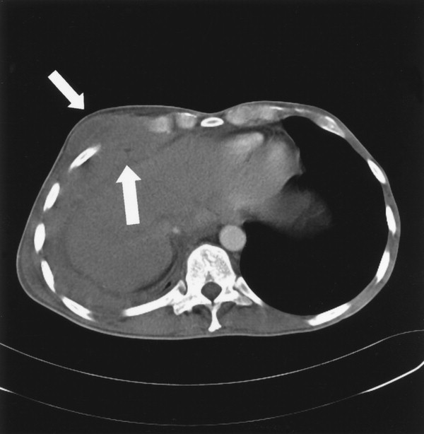
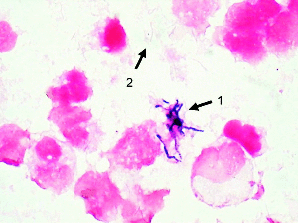
A 43 YOM had painful swelling of right chest


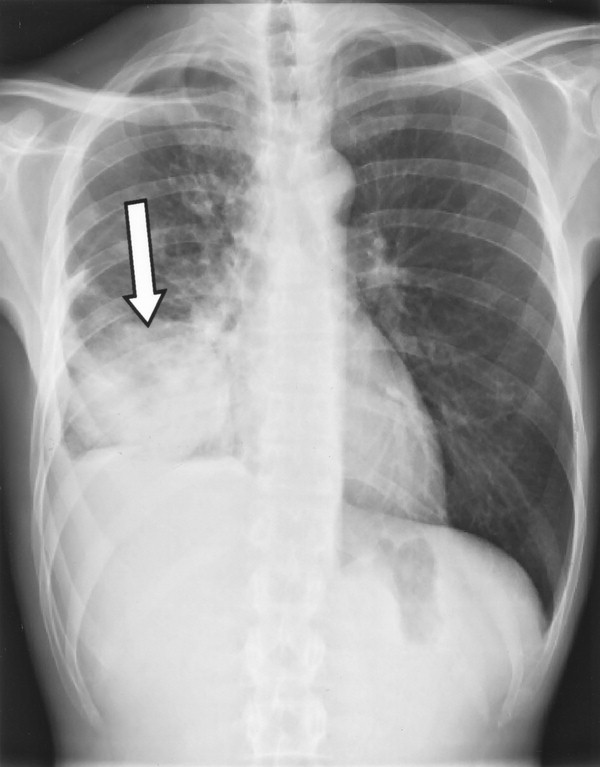
   ชายอายุ 43 ปีถูกส่งต่อมาเนื่องจากเจ็บหน้าอก ไอมีเสมหะและภาพรังสีทรวงอกมีความผิดปกติดังรูป 1 ผู้ป่วยมีประวัติติดเสพติด แต่ไม่มีโรคประจำตัวอื่นๆ แพทย์จากรพ.เดิมคิดถึงวัณโรคปอดแต่ยังไม่สามารถพิสูจน์ได้

PE: moderately ill, generalized wasting of body muscle and subcutaneous fat. Body weight was 48 kg (body mass index, 16.1). low-grade fever and tachycardia. The oral cavity showed dental caries. There was no local or generalized lymphadenopathy. A slightly painful swelling was palpable in the right sixth intercostal space. Percussion of the right hemithorax was dull and tender. Diminished breathing sounds were heard on auscultation, without crackles or rales. The abdomen was not painful, and there was no organomegaly.

Laboratory tests: WBC 26000/cumm with normal differentiation, anemia (hemoglobin level, 5.2 mmol/L), thrombocytosis (platelet count, 819000/mL), and an elevated C-reactive protein level (200 mg/L).
Liver enzyme levels and kidney function tests: normal.

Bronchoscopy: no endobronchial abnormalities, and analysis of bronchoalveolar lavage fluid and transbronchial lung biopsy samples did not yield a diagnosis.


Posted by : chpantip , E-mail : (chpantip@medicine.psu.ac.th) ,
Date : 2008-11-17 , Time : 12:58:20 , From IP : 172.29.3.68


ความคิดเห็นที่ : 1


   รูป 1 คือ CXR
รูป 2 คือ CT of the thorax
พบความผิดปกติอะไรบ้างคะ คิดถึงโรคอะไรบ้าง

Bronchoscopy ไม่พบความผิดปกติ จะทำอะไรต่อดี


Posted by : chpantip , E-mail : (chpantip@medicine.psu.ac.th) ,
Date : 2008-11-17 , Time : 13:03:34 , From IP : 172.29.3.68


ความคิดเห็นที่ : 2


   Problem lists
1.Low-grade fever
2.Malnutrition
3.Mass at Lt.lung
4.WBC 26000/cumm with normal differentiation, anemia (hemoglobin level, 5.2 mmol/L), thrombocytosis (platelet count, 819000/mL), and an elevated C-reactive protein level (200 mg/L).

-Record V/S , observe fever
-try feed ร่วมกับให้ 5%DN/2 IV
-เจาะ aspirate ที่ mass ส่งย้อม gram-stain
-ทำ Percutaneous biopsy ที่บริเวณ mass ที่ผ่าน abdominal wall ขึ้นมา (จากรูปที่2)
-ให้ Pen V 2 ล้านยูนิต ไปก่อน ก่อนให้อย่าลืมทำ skin test ด้วยครับ


Posted by : harder , Date : 2008-11-17 , Time : 15:33:51 , From IP : 172.29.22.43

ความคิดเห็นที่ : 3


   ขอบคุณคุณหมอ harder มากค่ะ คุณหมอตอบได้ถูกต้องแล้วค่ะ

CT of the thorax (figure 2) showed a mass protruding through the thoracic wall.

เจาะที่ตำแหน่งที่บวมนูน ได้หนอง
ย้อมหนองด้วย gram stain พบเชื้อดังในรูป 3 เป็นเชื้อ 2 ชนิด คิดว่าเป็นเชื้ออะไรบ้างคะ


Posted by : chpantip , E-mail : (chpantip@medicine.psu.ac.th) ,
Date : 2008-11-18 , Time : 11:02:10 , From IP : 172.29.3.68


ความคิดเห็นที่ : 4


   ลูกศร 1 : Gram positive rod คิดว่าเป็นพวก Actinomyces ครับ
ลูกศร 2 : เส้นเล็กนิดเดียว แต่คิดว่าเป็น Gram negative rod ครับ แต่นึกเชื้อไม่ออกครับ ว่าควรเป็นเชื้ออะไรมากที่สุด รอความเห็นจากท่านอื่นล่ะกันครับ

ปล.พี่ Sagittarius , Aomnaka , arlim ไปไหนแล้วอ่าครับ ช่วงนี้เงียบไปเลย


Posted by : harder , Date : 2008-11-18 , Time : 13:28:04 , From IP : 172.29.22.43

ความคิดเห็นที่ : 5


   คุณหมอ Harder ตอบได้ถูกต้องแล้วค่ะ ดิฉันขอเฉลยนะคะ

Figure 1. Chest radiograph demonstrating a shadow on the lower field of the right lung (arrow) and pleural thickening.

Figure 2. CT scan of the thorax, showing a mass protruding through the thoracic wall (arrows)

Figure 3. Gram stain of the purulent transthoracic aspirate, showing irregular gram-positive rods without branching (arrow 1) and slender, pointed gram-negative rods (arrow 2) (oil immersion)

ได้ส่งหนองที่เจาะได้ไปเพาะเชื้อ aerobic และ anaerobic cultures ขึ้นเชื้อแบคทีเรีย 3 strain
1. Anaerobic, slow-growing, catalase-negative gram-positive rod—was identified as Actinomyces israelii by 16S RNA gene sequencing.
2. Anaerobic, very slow–growing, slender gram-negative rod with pointed ends, identified by 16S RNA gene sequencing as Fusobacterium nucleatum.
3. Fastidious, catalase-positive gram-negative rod that was identified as Actinobacillus actinomycetemcomitans.


Posted by : chpantip , E-mail : (chpantip@medicine.psu.ac.th) ,
Date : 2008-11-19 , Time : 11:47:23 , From IP : 172.29.3.68


ความคิดเห็นที่ : 6


   พบได้บ่อยๆ ว่ามีหลาย species ของ bacteria ใน clinical sample ขึ้นร่วมกับ Actinomyces species ในการเพาะเชื้อ โดยที่เชื้อเหล่านี้เป็น flora ที่จะบอกถึงแหล่งของการติดเชื้อ ดังเช่นในรายนี้เกิดจากเชื้อในช่องปากมากที่สุด การมี dental hygiene ไม่ดีเป็น predisposing factor ของการเกิด thoracic actinomycosis ทางที่เกิดการติดเชื้ออาจเป็นจาก aspiration หรือทาง deep cervical fascial space ไปสู่ mediastinum

ปัจจุบัน เป็นที่ยอมรับกันแล้วว่า actinomycotic infections เป็นการติดเชื้อของเชื้อหลายชนิดและเสริมฤทธิ์กัน แต่ monomicrobic infection จาก Actinomyces species ก็เกิดขึ้นได้

พบได้บ่อยว่าเพาะเชื้อขึ้น Actinobacillus actinomycetemcomitans, Eikenella corrodens, Fusobacterium species, Bacteroides species, Capnocytophaga species, Staphylococcus species, Streptococcus species, และ Enterobacteriaceae ร่วมกับ Actinomyces species โดยขึ้นอยู่กับตำแหน่งของการติดเชื้อ เชื้อที่พบร่วมกันนี้ช่วยเสริมการเกิด actinomycotic lesion ดังนั้นจึงควรหาว่ามี potential copathogens หรือไม่เพื่อให้การรักษาที่เหมาะสม (ตัวอย่างเช่น A. actinomycetemcomitans สัมพันธ์กับการติดเชื้อที่เรื้อรังและการรักษาที่ล้มเหลวสามารถทำให้เชื้อคงอยู่ได้


Posted by : chpantip , E-mail : (chpantip@medicine.psu.ac.th) ,
Date : 2008-11-27 , Time : 12:06:29 , From IP : 172.29.3.68


สงวนสิทธิ์การแสดงความคิดเห็นสำหรับ สมาชิกเท่านั้น