ความคิดเห็นทั้งหมด : 4

ชายอายุ 24 ปี ปวดข้อเท้าและข้อที่มือทั้ง 2 ข้าง


   ชายอายุ 24 ปีมาตรวจด้วยเรื่องปวดข้อเท้าและข้อเล็กๆ ที่มือทั้ง 2 ข้างโดยไม่มีบวม

โปรดอ่าน image study ของผู้ป่วยและให้การวินิจฉัยโรคด้วยค่ะ

รูป A คือ lateral film of the skull
รูป B คือ sagittal view of a T1-weighted magnetic resonance image of the skull
รูป C คือ radiograph of the tibia and fibula


Posted by : chpantip , E-mail : (chpantip@medicine.psu.ac.th) ,
Date : 2008-11-12 , Time : 11:05:55 , From IP : 172.29.3.68


ความคิดเห็นที่ : 1


   อ่านไม่ถูกเหมือนกันคับ แหะแหะ แต่มีปวดข้อเท้าและข้อเล็กๆ ไม่บวม แสดงว่าไม่น่าจะมี inflammation อาจจะเกิดที่กระดูกโดยตรง เดาว่า Hyperparathyroidism ล่ะกันครับ เพราะรู้สึกกระดูกจะบางๆครับ (รึเปล่าหว่า?) ซึ่งอาจทำให้เกิดอาการเจ็บที่กระดูกได้ครับ

แต่ผู้ป่วยน่าจะมาด้วยอาการเพลีย อาเจียน ดื่มน้ำและปัสสาวะมาก มากกว่าอ่ะครับ

แล้วจะรอเฉลยครับผม


Posted by : harder , Date : 2008-11-12 , Time : 13:51:12 , From IP : 172.29.22.43

ความคิดเห็นที่ : 2


   Imaging study ของผู้ป่วยรายนี้
รูป A : Hair-on-end appearance of the outer table of skull (spare occiput)
รูป B : expansion of the diploetic portion of the cranium
รูป C: osteopenia, widening of marrow cavity and cortical thinning
แสดงถึง hyperplasia of bone marrow เข้าได้กับ thalassemia major


Posted by : lille , Date : 2008-11-12 , Time : 19:05:07 , From IP : 172.29.4.2

ความคิดเห็นที่ : 3


   คุณ lille เก่งจังครับ พอเห็นเฉลย เลยพอนึกออกครับ พออ่านฟิล์มทีไร ผมเน่าทุกที อ่านไม่ค่อยได้เลยครับ อาจารย์ก็สอนหลายครั้งแล้ว แต่อ่านให้ได้อย่างนี้ไม่ได้ซักที

ถ้าคุณ lille มีข้อแนะนำเรื่องการอ่านฟิล์ม โปรดชี้แนะด้วยนะครับ ขอบคุณครับ


Posted by : harder , Date : 2008-11-12 , Time : 20:46:07 , From IP : 172.29.22.43

ความคิดเห็นที่ : 4


   คุณหมอ lille เก่งมากๆเลยค่ะ อีกหน่อยคุณหมอ harder ก็จะเก่งมากเหมือนกันค่ะ

ขอเพิ่มเติมข้อมูลนะคะ

ผู้ป่วยเคยได้รับเลือดมา 37 ครั้ง เขามี fetal hemoglobin value = 94%และ A2 hemoglobin value =1 %
พ่อของผู้ป่วยมี A2 hemoglobin percent สูง
แม่ของผู้ป่วยมี fetal hemoglobin percent สูงและ A2 hemoglobin value ปกติ

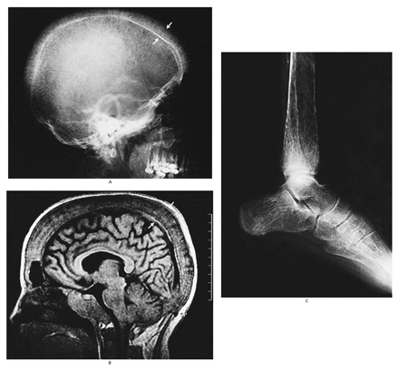
รูป A lateral film of the skull: generalized osteopenia, widening of the diploetic space (arrows), and thinning of the outer table. The characteristic "hair-on-end" appearance of the outer table spares the inferior aspect of the occiput.

รูป B sagittal view of a T1-weighted magnetic resonance image of the skull: expansion of the diploetic portion of the cranium (arrows) and the clivus and obliteration of the air space of the sphenoidal sinus.

รูป C radiograph of the tibia and fibula: coarse osteopenia, cortical thinning, small cystic lesions, and a widened marrow cavity.

ลักษณะทั้งหมดนี้เข้าได้กับการมี massive hyperplasia ของ bone marrow
การวินิจฉัยโรค: thalassemia major


Posted by : chpantip , E-mail : (chpantip@medicine.psu.ac.th) ,
Date : 2008-11-13 , Time : 11:47:36 , From IP : 172.29.3.68


สงวนสิทธิ์การแสดงความคิดเห็นสำหรับ สมาชิกเท่านั้น