ความคิดเห็นทั้งหมด : 7

ชาย 64 ปี มีไข้และจมูกบวมและเจ็บมา 5 วัน


   ชายอายุ 64 ปี มาที่ ER ด้วยไข้ ร่วมกับมีจมูกบวมและเจ็บมา 5 วัน ผู้ป่วยเป็น COPD อยู่เดิม

PE: T 39°C, RR 22/min, BP 120/70 mm Hg, HR 86/min
chest auscultation: left sided crackles were heard on
He had a painful fluctuant abscess of the anterior nose.

Laboratory tests
CBC: WBC 38000/cumm with 49% band forms
C-reactive protein (350 mg/L, normal range <10 mg/L).
Chest radiography on admission showed an infiltrate of the left upper lung.

1. บอกปัญหาของผู้ป่วย
2. บอกการวินิจฉัยที่น่าจะเป็นมากที่สุดรวมถึงเชื้อก่อโรคด้วย
3. ท่านจะ manage ผู้ป่วยอย่างไรต่อไป


Posted by : chpantip , E-mail : (chpantip@medicine.psu.ac.th) ,
Date : 2008-09-05 , Time : 12:51:19 , From IP : 172.29.3.68


ความคิดเห็นที่ : 1


   1.
- sepsis
- soft tissue infection at nose (ไม่เห็นรูป) น่าจะเป็น abscess
- Left upper lobe pneumonia
- Host COPD and elderly

2.
ให้คิดเป็นเรื่องเดียวกัน
- Bacterial pneumonia and abscess at nose

3.
- management sepsis ดูเรื่อง volume status and assessment , organ dysfunction
- G/S , H/C and pus C/S
- appropriated ATB cover organism ; staphylococcus streptococcus and anaerobe
choose ATB Augmantin IV หรือ Cloxacillin+Clindamycin IV
- Local control ; debridement at abscess
- F/U clinical ต่างๆ ระวัง complication ของ pneumonia

ขอบคุณครับ


Posted by : sagittareusinternist , Date : 2008-09-05 , Time : 15:09:45 , From IP : 172.29.5.213

ความคิดเห็นที่ : 2


   ไม่ทราบว่าอาจเป็น Diffuse panbronchiolitis ได้ไม๊ครับ แค่เสนอ DDx เฉยๆครับ เพราะ DPB ผู้ป่วยมักไม่สูบบหุรี่ แต่ในเคสนี้ผู้ป่วยเป็น COPD ซึ่งกว่า 90% เกิดจากสูบบุหรี่

การรักษาผมคิดว่าย้อม gram stain ให้ ATB ที่ครอบคลุม แล้วก็ส่ง culture ดูเชื้อจำเพาะและความไวต่อการดื้อยาอ่าครับ

ยังเป็นนศพ.ฝากเนื้อฝากตัวด้วยครับ


Posted by : harder , Date : 2008-09-06 , Time : 18:21:41 , From IP : 172.29.22.43

ความคิดเห็นที่ : 3


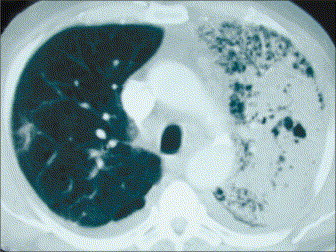
   ขอบคุณคุณหมอ sagittareusinternist และนศพ.harder มากค่ะ ยินดีต้อนรับสู่ e-consult ค่ะ

แพทย์ได้เจาะและระบายหนองฝีที่จมูกพร้อมให้ยา amoxicillin/clavulanic acid, and amikacin ผลการเพาะเชื้อจากเลือดและหนองจากจมูกขึ้น methicillin-susceptible Staphylococcus aureus
ในช่วงสัปดาห์แรกของการรักษา ผู้ป่วยหอบเหนื่อยมากขึ้น
ได้ทำ Chest CT วันที่ 6 ของการรักษา
ถาม
1. พบความผิดปกติอะไรจาก Chest CT บ้าง
2. ท่านจะส่ง work up อะไรบ้างเพื่อให้ทราบถึง virulence factor ของเชื้อนี้


Posted by : chpantip , E-mail : (chpantip@medicine.psu.ac.th) ,
Date : 2008-09-10 , Time : 15:35:46 , From IP : 172.29.3.68


ความคิดเห็นที่ : 4


   1.พบความผิดปกติอะไรจาก Chest CT บ้าง
- ปอดด้านซ้ายมีพยาธิสภาพลักษณะเหมือน Fluid-density กระจายไปทั่วปอด
- ปอดด้านขวามี Fluid-density เล็กน้อยที่ขอบด้านนอกของปอด
2.ท่านจะส่ง work-up อะไรบ้างเพื่อให้ทราบถึง virulence factor ของเชื้อนี้
- hyaluronidase, protease, coagulase, lipases, deoxyribonucleases และ enterotoxin

ตอบถูก-ผิดประการใด ช่วยสั่งสอนด้วยครับ


Posted by : harder , Date : 2008-09-12 , Time : 15:57:01 , From IP : 172.29.22.43

ความคิดเห็นที่ : 5


   ขอบคุณคุณหมอ Harder มากค่ะ

ตอบ
Chest CT วันที่ 6 พบ multiple confluent lesions bilaterally with abscess formations and mediastinal lymphadenopathy

เนื่องจากผู้ป่วยมี necrotizing pneumonia จึงนึกถึงว่า เชื้อก่อโรคน่าจะเป็น S. aureus สายพันธุ์ที่สร้าง Panton-Valentine leucocidin (PVL) ได้ส่ง PCR และตรวจพบ PVL loci (lukS-PV and lukF-PV)

ผู้ป่วยได้รับการรักษาด้วย IV cloxacillin นาน 4 สัปดาห์ ตามด้วย clindamycin กินนาน 4 สัปดาห์จนฝีที่ปอดหายไปหมด จากการติดตามหลังสุดที่ 3 เดือนต่อมา ผู้ป่วยสบายดี โดยที่ยังคงมี perforated nasal septum

ขอถามต่อนะคะ

Panton-Valentine leucocidin (PVL) มีความสำคัญอย่างไร


Posted by : chpantip , E-mail : (chpantip@medicine.psu.ac.th) ,
Date : 2008-09-15 , Time : 16:09:35 , From IP : 172.29.3.68


ความคิดเห็นที่ : 6


   Panton-Valentine leukocidin (PVL) เป็น cytotoxin ของ S.aureus ที่ทำให้เกิดการทำลายเนื้อเยื่อปอดและ host defence cell เช่น WBC , Macrophage หรือ monocyte
ซึ่งจะส่งผลทำให้เกิดโรคที่เรียกว่า Necrotizing pneumonia


Posted by : harder , Date : 2008-09-15 , Time : 21:14:00 , From IP : 172.29.22.43

ความคิดเห็นที่ : 7


   ขอบคุณคุณหมอ harder มากค่ะ คุณหมอตอบถูกต้องแล้วค่ะ ดิฉันขอขยายความนะคะ

S. aureus เป็นเชื้อก่อโรคประมาณ 2% ของ community-acquired pneumonia และ10% ของ nosocomial pneumonia โดยมีอัตราตายจาก 30% ถึง 80% ในรายที่มีbacteremia การมี nasal colonisation ด้วย S. aureus เป็นปัจจัยเสี่ยงที่สำคัญมากของ invasive infection ที่เกิดตามมา ในยุโรป พบว่ามากกว่า 20% ของผู้ป่วยทั้งหมดที่รับการรักษาใน non-surgical ward เป็น nasal carrier ของ S. aureus และ 80% ของ bacteremic infection ใน carrier สัมพันธ์กับการมีเชื้อนี้ colonize อยู่ในช่องจมูกส่วนหน้า

PVL เป็น virulence factor ชนิดหนึ่งของ S. aureus ซึ่งสามารถเปลี่ยนแปลง permeability ของ membrane ของ human polymorphonuclear neutrophil และทำให้เซลล์ตาย proinflammatory effect ของ PVL และ lytic activity ต่อ granulocyte ทำให้เกิด necrosis จึงทำให้เกิด rapidly progressive necrotising pneumonia และ skin and soft tissue infections รวมถึง necrotising fasciitis

การปรากฏอย่างรวดเร็วของ community-acquired strain ของ methicillin-resistant S aureus (MRSA) ในต่างประเทศในทศวรรษที่ผ่านมาทำให้มีความสนใจ PVL-associated disease มากขึ้น เนื่องจาก 70% ของ community-acquired MRSA strain มี PVL genes ส่วน nosocomial S aureus strain มี PVL น้อยกว่า 5% community-acquired MSSA strain ก็สามารถมี PVL genes ได้เช่นกัน MSSA isolate ที่มี lukS-PV and lukF-PV อาจทำให้เกิด 70% ของ recurrent skin abscesses หรือ furuncles และมากกว่า 90% ของnecrotising pneumonia ที่เกิดจาก S aureus PVL สัมพันธ์อย่างมากกับ necrotic lesion ที่เป็นกับผิวหนังหรือเยื่อเมือก PVL-positive S aureus necrotising pneumonia พบได้บ่อยกว่าในเด็กเล็กที่มีภูมิต้านทานปกติหรือผู้ใหญ่ที่มี influenza-like syndrome นำมาก่อน การมี PVL สัมพันธ์กับความรุนแรงของโรค ตั้งแต่ cutaneous infection ที่ต้องการ surgical drainage จนถึง necrotising pneumonia โดยมีอัตราตายสูงถึง 75% และมีการตายที่รวดเร็วภายใน 48 ชั่วโมงของการรับเข้ารักษาในรพ.

รูปและเรื่อง จาก Laifer G. Lancet 2006;367:1628


Posted by : chpantip , E-mail : (chpantip@medicine.psu.ac.th) ,
Date : 2008-09-22 , Time : 13:01:57 , From IP : 172.29.3.68


สงวนสิทธิ์การแสดงความคิดเห็นสำหรับ สมาชิกเท่านั้น