ขอเฉลยค่ะ
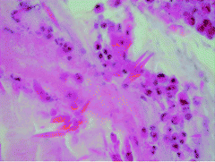
รูป C, pathological examination of the bronchial mass showed mucin containing numerous eosinophils as well as occasional Charcot leyden crystals;
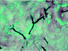
รูป D, Gomori methenamine silver stain highlights occasional fungal hyphae morphologically consistent with Aspergillus species (มี dichotomous branching หรือ acute angle branching คือ แตกกิ่งแหมือนกิ่งไม้ )
Diagnosis: Allergic bronchopulmonary aspergillosis (ABPA)
ABPA เป็นโรคทางระบบหายใจที่พบไม่บ่อยแต่มีความรุนแรง ลักษณะจำเพาะของโรคนี้คือ การอักเสบและการอุดตันของหลอดลมอย่างเรื้อรังจากการมี persistent colonization โดย Aspergillus fumigatus และเกิดการแพ้เชื้อนี้
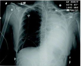
อาการโรคที่สำคัญ หอบหืด ที่มีภาวะแทรกซ้อนเป็นๆหายๆ จากการอุดตันของหลอดลม ไข้ อ่อนเพลีย ไอมีเสมหะเหนียวสีน้ำตาล และมี peripheral blood eosinophilia ABPA สามารถมี progressive airflow obstruction และ pulmonary fibrosis ตามมาและนำไปสู่ความพิการที่เรื้อรังและรุนแรงได้
การวินิจฉัย ABPA อาศัย 7 primary diagnostic criteria:
1. episodic bronchial obstruction (asthma),
2. peripheral eosinophilia,
3. immediate scratch test reactivity to Aspergillus antigen,
4. precipitating antibodies to Aspergillus antigen,
5. elevated serum IgE concentrations,
6. history of pulmonary infiltrates (transient or fixed), and
7. central bronchiectasis.
Secondary diagnostic criteria
1. repeated detection of Aspergillus species in sputum samples using stain and/or culture,
2. a history of expectoration of brown mucous plugs or flecks,
3. elevated specific IgE concentration directed against Aspergillus
antigen, and
4. Arthus reaction (late skin reactivity) to Aspergillus antigen.
ABPA อาจมีความรุนแรงน้อย แบบ acute corticosteroid-responsive asthma จนเป็นรุนแรงถึงขนาด corticosteroid-dependent asthma และเป็น fibrotic end-stage lung disease with honeycombed lung ถ้าสามารถให้การวินิจฉัยและรักษาได้ตั้งแต่แรกก็จะสามารถลด disability จาก ABPA ได้
รักษา ABPA ด้วย corticosteroids และยาต้านเชื้อรา คือ azoles รูปยากิน
Systemic corticosteroids เป็นการรักษาหลักของ ABPA โดย corticosteroids ไปยับยั้ง inflammatory response ที่เกิดชึ้นจาก A. fumigates อาจต้องให้ oral corticosteroids ขนาดสูงเป็นเวลานาน ซึ่งจะช่วยควบคุม asthma และเร่งให้ pulmonary infiltrates หายเร็วขึ้น
A. fumigatus ทำให้เกิด persistent colonization ใน ABPA แต่ไม่ทำให้เกิด invasive disease การกำจัดเชื้อนี้ อาจลดความรุนแรงของโรค meta-analysis ฉบับหนึ่งแสดงให้เห็นว่าการให้ itraconazole ร่วมด้วย ทำให้ serum IgE level (marker of immune activation ตัวหนึ่ง) ลดลงมากกว่า 25% (OR compared with placebo, 3.26; range, 1.30–8.15)
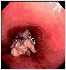
ในผู้ป่วยรายนี้ ผลการย้อมและเพาะเชื้อพบว่าเป็น A. fumigatus การเอาก้อนออกทำให้ปอดขยายได้เกือบทั้งหมด การตรวจทางพยาธิวิทยาของก้อนในหลอดลม พบ mucin ที่มี eosinophil เป็นจำนวนมาก และมี Charcot leyden crystals ด้วย Gomori methenamine silver stain พบ fungal hyphae ของเชื้อราที่มีลักษณะเข้าได้กับ Aspergillus species (รูป C) ผู้ป่วยมี eosinophilia ระดับ total IgE และ serum-specific IgE for A. fumigatus ขึ้นสูงทั้งคู่ เขาหายดีจากการรักษาด้วย corticosteroid และ voriconazole
เรื่อและรูปจาก Ali Hassoun. Cauliflower Mass Obstructing the Left Main-Stem Bronchus. Clin Infect Dis 2008;47:540–1.
Posted by : chpantip , E-mail : (chpantip@medicine.psu.ac.th) ,
Date : 2008-08-17 , Time : 14:08:33 , From IP : 172.29.3.68
|