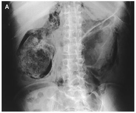
หญิง 50 ปี ซึมลงและมีความดันเลือดต่ำหญิงอายุ 50 ปี ญาตินำส่งมาที่ ER เนื่องจากซึมลงและมีความดันเลือดต่ำ ผู้ป่วยมีไข้ต่ำๆมานาน 1 เดือน ก่อนหน้านี้ผู้ป่วยสบายดี PE: an immobile, tender, firm mass was palpated to the right of the umbilicus. Lab: white-cell count 17,200 /cumm, serum glucose level of 607 mg/dl, BUN 70 mg/dl, serum creatinine 4.0 mg/dl. 1. abdominal radiograph ดังในรูป มีความผิดปกติอะไรบ้าง 2. จงบอกการวินิจฉัยโรค และ management Posted by : chpantip , E-mail : (chpantip@medicine.psu.ac.th) , Date : 2008-08-10 , Time : 16:33:35 , From IP : 172.29.3.68 |
1. Heterogenous mass ที่ด้านท้องด้านขวา และรอบๆ mass มี air อยู่ล้อมรอบ ลักษณะของ mass เหมือนมี capsule หุ้มอยู่ค่ะ และตรงกลางที่ติดกับ spine ดูหนาขึ้น คิดว่าเป็นขั้วไตข้างขวาค่ะ ส่วนด้านซ้ายมีลักษณะเป็นเส้นขาวๆเป็นทางยาว คิดว่าเป็น vessele ค่ะ 2. จากประวัติ ตรวจร่างกาย ผล x-ray และ lab ผู้ป่วยรายนี้มีปัญหา - prolong fever - alteration of consciousness with shock ??(ไม่มีข้อมูล V/S ค่ะ แต่ BP ต่ำ คิดว่าน่าจะเริ่มมี shock แล้ว) - leukocytosis - Hyperglycemia - renal failure - abdominal mass คิดว่าสาเหตุที่ทำให้เกิดปัญหาต่างๆเหล่านี้เกิดจาก malignancy cause ค่ะ และที่คิดถึงคือ RCC ค่ะ management - IV fluid resuscitate with NSS - Empirical ATB with Ceftriaxone ค่ะ เพราะคิดว่าผู้ป่วยน่าจะมี infection ร่วมด้วย และสาเหตุที่ทำให้เกิด alteration of conscious ยังไม่สามารถแยกได้ว่าเกิดจาก hyperglycemia หรือ RF หรือ sepsis อย่างใดอย่างหนึ่งค่ะ - แก้ไขภาวะ hyperglycemia --> RI iv ค่ะ - metabolic และ septic work up ค่ะ และคิดว่าจำเป็นต้องทำ CT abdomen เพื่อดู abdominal mass ขอบเขตของ mass metastasis และ LN ในท้องค่ะ Posted by : nongnham , E-mail : (nongnham025@gmail.com) , Date : 2008-08-13 , Time : 11:37:00 , From IP : 222.123.181.146 |
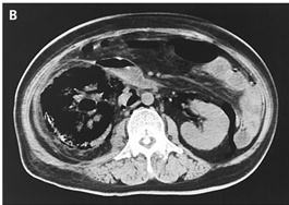
ขอบคุณคุณหมอ Nongnham มากค่ะ ตอบถูกต้องแล้วค่ะ ที่ให้ management แบบ septic shock Abdominal radiograph: gas throughout the right kidney Computed tomography (CT) of the abdomen: extensive destruction of the right renal parenchyma with associated gas, as well as gas in the retroperitoneal tissues. ผู้ป่วยมีเบาหวาน (serum glucose level แรกรับ = 607 mg /dl และ HbA1C = 12.2%). ผู้ป่วยไม่ได้มี diabetic ketoacidosis. การวินิจฉัยโรค emphysematous pyelonephritis ในผู้ป่วยเบาหวาน ผู้ป่วยได้รับการผ่าตัด right nephrectomy อย่างเร่งรีบ ผลการเพาะเชื้อจากหนองที่ไตและจากเลือดขึ้น E. coli. ผู้ป่วยมีอาการดีขึ้นและหายโดย serum creatinine กลับเป็นปกติ เธอได้รับการรักษาเบาหวานด้วย insulin และสบายดีหลังจากนั้น เรื่องและรูปจาก Tajima K., Kurabayashi M. N Engl J Med 2006;355:2467. Emphysematous pyelonephritis พบได้น้อยแต่เป็น potentially life-threatening disease มีลักษณะที่สำคัญคือ มี gas accumulation อยู่ในเนื้อไตหรือรอบไต ผู้ที่เป็นโรคนี้ส่วนใหญ่เป็นเบาหวาน มาด้วยอาการไข้และปวดเอว เชื้อก่อโรคที่พบบ่อยที่สุดคือ E. coli ตามมาด้วย Klebsiella pneumoniae การวินิจฉัย emphysematous pyelonephritis ทำได้ยาก ต้องใช้ imaging studies การวินิจฉัยที่ล่าช้าจะเพิ่ม morbidity และ mortality CT scan เป็น imaging modality สำหรับการวินิจฉัยที่ดีที่สุด เพราะจะเห็น air ใน renal parenchyma หรือ perirenal space abdominal radiograph ก็ช่วย แต่ sensitivity ต่ำกว่า CT scan การรักษา emphysematous pyelonephritis : ให้ยาปฏิชีวนะ ร่วมกับ adequate drainage Selective nephrectomy ควรพิจารณาทำในรายที่ high-risk ซึ่งมี shock, thrombocytopenia, acute renal failure, หรือ disturbed consciousness Posted by : chpantip , E-mail : (chpantip@medicine.psu.ac.th) , Date : 2008-08-13 , Time : 14:24:41 , From IP : 172.29.3.68 |